Generic selectors
Exact matches only
Search in title
Search in content
Post Type Selectors
Search in posts
Search in pages
Filter by Categories
15th National Conference of the IAOMFP, Chennai, 2006
Abstract
Abstracts from current literature
Acne in India: Guidelines for management - IAA Consensus Document
Addendum
Announcement
Art & Psychiatry
Article
Articles
Association Activities
Association Notes
Award Article
Book Review
Brief Report
Case Analysis
Case Letter
Case Letters
Case Notes
Case Report
Case Reports
Clinical and Laboratory Investigations
Clinical Article
Clinical Studies
Clinical Study
Commentary
Conference Oration
Conference Summary
Continuing Medical Education
Correspondence
Corrigendum
Cosmetic Dermatology
Cosmetology
Current Best Evidence
Current Issue
Current View
Derma Quest
Dermato Surgery
Dermatopathology
Dermatosurgery Specials
Dispensing Pearl
Do you know?
Drug Dialogues
e-IJDVL
Editor Speaks
Editorial
Editorial Remarks
Editorial Report
Editorial Report - 2007
Editorial report for 2004-2005
Errata
Erratum
Focus
Fourth All India Conference Programme
From Our Book Shelf
From the Desk of Chief Editor
General
Get Set for Net
Get set for the net
Guest Article
Guest Editorial
History
How I Manage?
IADVL Announcement
IADVL Announcements
IJDVL Awards
IJDVL AWARDS 2015
IJDVL Awards 2018
IJDVL Awards 2019
IJDVL Awards 2020
IJDVL International Awards 2018
Images in Clinical Practice
In Memorium
Inaugural Address
Index
Knowledge From World Contemporaries
Leprosy Section
Letter in Response to Previous Publication
Letter to Editor
Letter to the Editor
Letter to the Editor - Case Letter
Letter to the Editor - Letter in Response to Published Article
LETTER TO THE EDITOR - LETTERS IN RESPONSE TO PUBLISHED ARTICLES
Letter to the Editor - Observation Letter
Letter to the Editor - Study Letter
Letter to the Editor - Therapy Letter
Letter to the Editor: Articles in Response to Previously Published Articles
Letters in Response to Previous Publication
Letters to the Editor
Letters to the Editor - Letter in Response to Previously Published Articles
Letters to the Editor: Case Letters
Letters to the Editor: Letters in Response to Previously Published Articles
Medicolegal Window
Messages
Miscellaneous Letter
Musings
Net Case
Net case report
Net Image
Net Images
Net Letter
Net Quiz
Net Study
New Preparations
News
News & Views
Obituary
Observation Letter
Observation Letters
Oration
Original Article
ORIGINAL CONTRIBUTION
Original Contributions
Pattern of Skin Diseases
Pearls
Pediatric Dermatology
Pediatric Rounds
Perspective
Presedential Address
Presidential Address
Presidents Remarks
Quiz
Recommendations
Regret
Report
Report of chief editor
Report of Hon : Treasurer IADVL
Report of Hon. General Secretary IADVL
Research Methdology
Research Methodology
Resident page
Resident's Page
Resident’s Page
Residents' Corner
Residents' Corner
Residents' Page
Retraction
Review
Review Article
Review Articles
Reviewers 2022
Revision Corner
Self Assessment Programme
SEMINAR
Seminar: Chronic Arsenicosis in India
Seminar: HIV Infection
Short Communication
Short Communications
Short Report
Snippets
Special Article
Specialty Interface
Studies
Study Letter
Study Letters
Supplement-Photoprotection
Supplement-Psoriasis
Symposium - Contact Dermatitis
Symposium - Lasers
Symposium - Pediatric Dermatoses
Symposium - Psoriasis
Symposium - Vesicobullous Disorders
SYMPOSIUM - VITILIGO
Symposium Aesthetic Surgery
Symposium Dermatopathology
Symposium-Hair Disorders
Symposium-Nails Part I
Symposium-Nails-Part II
Systematic Review and Meta-Analysis
Systematic Reviews and Meta-analyses
Systematic Reviews and Meta-analysis
Tables
Technology
Therapeutic Guideline-IADVL
Therapeutic Guidelines
Therapeutic Guidelines - IADVL
Therapeutics
Therapy
Therapy Letter
Therapy Letters
View Point
Viewpoint
What’s new in Dermatology
View/Download PDF

Translate this page into:

Letter to the Editor - Observation Letter
2020:86:5;568-570
doi: 10.4103/ijdvl.IJDVL_699_19
PMID: 32769307

The grid pattern of white superficial onychomycosis

Chander Grover1 , Deepak Jakhar1 , Sonal Sharma2
1 Department of Dermatology, University College of Medical Sciences and Guru Tegh Bahadur Hospital, New Delhi, India
2 Department of Pathology, University College of Medical Sciences and Guru Tegh Bahadur Hospital, New Delhi, India

Correspondence Address:
Chander Grover
Department of Dermatology, University College of Medical Sciences and Guru Tegh Bahadur Hospital, Dilshad Garden, New Delhi 110095
India
Published: 30-Jul-2020
How to cite this article:
Grover C, Jakhar D, Sharma S. The grid pattern of white superficial onychomycosis. Indian J Dermatol Venereol Leprol 2020;86:568-570
Copyright: (C)2020 Indian Journal of Dermatology, Venereology, and Leprology

Sir,

White superficial onychomycosis is a relatively uncommon type of onychomycosis where the fungus invades the superficial layers of the dorsal nail plate. The fungal colonies appear as white or yellow opacities on the nail plate surface producing “pseudo-leukonychia.” The clinical diagnosis is often missed and the dermoscopic features are not well-described due to paucity of such cases.[1]

A 38-year-old man presented with yellowish-white discoloration of all toenails for the past 1 year [Figure - 1]. On examination, the toenails had a friable looking nail plate surface with no evidence of subungual hyperkeratosis and periungual inflammation. Fingernails were spared. The rest of the mucocutaneous examination was noncontributory.

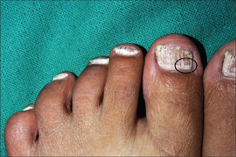
Figure 1: Clinical presentation of white superficial onychomycosis in a 38-year-old male. Note the involvement of multiple toe nails with white powdery deposits on the nail plate surface

Similarly, a 20-year-old woman presented with whitish discoloration of all toenails for the past 8 months. In this case also toenails appeared to have a friable nail plate surface with no signs of periungual inflammation.

Both cases were diagnosed clinically as white superficial onychomycosis, and onychoscopy was performed as the first step of assessment in both cases. We used a contact handheld dermatoscope (Dermlite DL4-3Gen; 10×; polarizing) and a universal serial bus dermatoscope (Dinolite AM7115MZT; 20–220×; polarizing) for the assessment. The purpose of using a universal serial bus dermatoscope was to observe any additional features at higher magnification (150×). Onychoscopy showed yellowish-white, opaque and friable debris on the nail plate, which was concentrated proximally [Figure - 2]. The appearance can be likened to “chickpea flour” sprayed on the nail unit (can be seen sometimes in housewives working with flour). On higher magnification (150×), these patches appeared to be composed of linear striae, progressing from the proximal to the distal end of the nail plate [Figure - 3]. In between, these patches showed a unique grid pattern within as well as between adjacent patches [Figure - 4]. Other features included nail plate discoloration, onychoschizia, loss of cuticle and inflamed nail folds. A 10% potassium hydroxide examination and histopathology [Figure - 5] confirmed the fungal etiology in both cases.

Figure 2: Onychoscopy of a case with white superficial onychomycosis. Whitish-yellowish opaque and friable looking debris (red arrow), predominantly concentrated on the proximal nail fold (Dermlite DL4-3Gen; ×100; polarizing)
Figure 3: Higher magnification showing linear striae (black arrow) progressing distally from the proximal debris, and nail plate discoloration (Dinolite AM7115MZT; ×150; polarizing)
Figure 4: Higher magnification showing the 'grid pattern' within as well as in-between the whitish patches (black circle). The cuticle is disrupted (Dinolite AM7115MZT; ×200; polarizing)
Figure 5: Photomicrograph of a nail plate biopsy showing fungal hyphae between the keratinocyte lamellae (red arrows) (PAS, ×400)

Onychoscopy has greatly improved the diagnostic armamentarium of dermatologists in diagnosing nail pathologies.[2],[3],[4],[5] Onychomycosis, especially the distal lateral subungual onychomycosis, has been extensively studied with onychoscopy. [Table - 1] shows the onychoscopic features of various types of onychomycosis. There is a paucity of literature for the uncommon forms such as endonyx and white superficial onychomycosis.[5],[6] Onychoscopy of the nail plate in the latter shows several small white or yellow opaque and friable patches.[5] Both cases in this report showed features similar to the previous report. In addition, we also noticed the proximal concentration of these friable patches, possibly due to the higher concentration of fungal elements near the nail folds and henceforth, the greater damage to the nail plate at this location. Higher magnification with the universal serial bus dermatoscope offered the visualization of a unique “grid pattern” within these friable patches. This grid pattern probably represents the macroview of the pattern of proliferation of fungal colonies on the nail plate surface. The most common differential diagnosis for the friable and brittle nail in such cases is nail lichen planus. Onychoscopy in nail lichen planus has been reported to show onychorrhexis, onychoschizia, pitting, chromonychia, pterygium, erythematous lunula, nail bed dyschromia, splinter hemorrhage and subungual hyperkeratosis.[7] Both onychopathies show typical onychoscopic features. Onychoscopy as the first step in diagnosis can help reduce the need for a biopsy.

Table 1: Various clinical types of onychomycosis and their onychoscopic features

In conclusion, a proximally concentrated yellowish white-colored friable, opaque debris on the nail plate and grid pattern of linear striae is a useful diagnostic clue for white superficial onychomycosis.

Declaration of patient consent

The authors certify that they have obtained all appropriate patient consent forms. In the form, the patients have given their consent for their images and other clinical information to be reported in the journal. The patients understand that their names and initials will not be published and due efforts will be made to conceal their identity, but anonymity cannot be guaranteed.

Financial support and sponsorship

Nil.

Conflicts of interest

There are no conflicts of interest.

References
1.
Yorulmaz A, Yalcin B. Dermoscopy as a first step in the diagnosis of onychomycosis. Postepy Dermatol Alergol 2018;35:251-8.
[Google Scholar]
2.
Piraccini BM, Alessandrini A, Starace M. Onychoscopy: Dermoscopy of the Nails. Dermatol Clin 2018;36:431-8.
[Google Scholar]
3.
Grover C, Jakhar D. Onychoscopy: A practical guide. Indian J Dermatol Venereol Leprol 2017;83:536-49.
[Google Scholar]
4.
Grover C, Jakhar D. Diagnostic utility of onychoscopy: Review of literature. Indian J Dermatopathol Diagn Dermatol 2017;4:31-40.
[Google Scholar]
5.
Alessandrini A, Starace M, Piraccini BM. Dermoscopy in the evaluation of nail disorders. Skin Appendage Disord 2017;3:70-82.
[Google Scholar]
6.
Jakhar D, Grover C, Kaur I. Dendritic pattern: Unique onychoscopic feature in endonyx. J Am Acad Dermatol 2019;81:e89-90.
[Google Scholar]
7.
Grover C, Kharghoria G, Bhattacharya SN. Linear nail bed dyschromia: A distinctive dermoscopic feature of nail lichen planus. Clin Exp Dermatol 2019;44:697-9.
[Google Scholar]

Fulltext Views
6,552

PDF downloads
1,007
Show Sections