Translate this page into:
"Pseudo" conditions in dermatology: Need to know both real and unreal
Correspondence Address:
Mohan H Kudur
Department of Dermatology and Venereology, Srinivas Institute of Medical Sciences and Research Centre, Mukka, Surathkal, Mangalore
India
| How to cite this article: Kudur MH, Hulmani M. "Pseudo" conditions in dermatology: Need to know both real and unreal. Indian J Dermatol Venereol Leprol 2012;78:763-773 |
Abstract
There are 'n' number of names and terminologies in dermatology. The real and unreal names lead to lot of confusion to the residents and practitioners of dermatology. The word 'pseudo' means 'unreal', 'false' or 'fake', and it has deep roots in dermatology providing herculean task to differentiate and understand the real conditions/diseases/signs in dermatology. We have made an attempt to list and describe the pseudo and associated real conditions in dermatology.Introduction
Studying and learning about true dermatological conditions is important before we actually treat patients, but, it is even more important to know what is untrue or unreal or pseudo. The pseudo conditions are different from the original diseases in etiology, treatment and prognosis. Pseudo conditions are look alike conditions occurring in the same site caused by different agents not related to the original cause. The well known pseudo conditions in dermatology are pseudo-scleroderma, pseudo-lymphoma, pseudo-bubo, pseudo Kaposi′s sarcoma, pseudo-ainhum, pseudo-Koebner′s phenomenon etc. We have classified pseudo conditions according to the major disease groups in dermatology.
Infections and Infestations
Folliculitis barbae and pseudo folliculitis barbae
Folliculitis barbae is a bacterial or fungal infecton of the hair follicle over the beard area [Figure - 1]. The bacterial folliculitis is caused by Staphylococcus aureus and it can be superficial or deep. The fungal folliculitis is caused by geophilic fungi and it will be of inflammatory type characterized by pustules over the beard area.
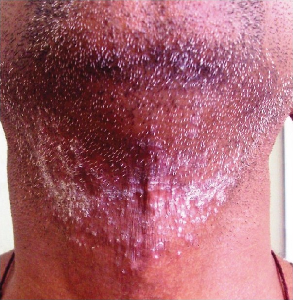 |
| Figure 1: Folliculitis: multiple pustules around hair follicles seen in beard area |
Pseudo-folliculitis barbae [1],[2] (Synonym: Pili incarnati)is not an infective condition, it is due to re-entry of sharp tips of cut hair in to skin. It is usually seen in beard area common in blacks and also seen in Indian patients. Close cutting of nasal hair results in pseudo folliculitis vibrissae.
Scabies and pseudo scabies
Scabies is caused by human mite (Sarcoptes scabiei var. hominis) with classical burrows involving finger web spaces [Figure - 2], axilla, breast, sides of abdomen, genitalia. Itching is severe and has a characteristic night aggravation. Scabies usually affects whole family, camps and hostels. The burrows are pathognomonic of scabies and the other common lesions seen are papules, excoriations and papulovesicles.
 |
| Figure 2: Scabies: multiple vesicles, pustules and papules seen in finger web spaces |
Pseudo scabies [3] is due to animal mite, commonly transmitted from dog. The clinical features are quite different like the itchy lesions are seen at sites of animal contact, sparing of finger web spaces and genitalia, lesions are more inflammatory, burrows are absent and the condition is self limiting.
Chancre redux and pseudo chancre redux
Chancre redux is relapse of primary chancre during the first two years of the disease (Relapsing stage of syphilis).
Pseudo chancre redux [4] is a term used to describe the appearance of solitary gumma at the site of original chancre.
Bubo and pseudo bubo
Bubo or inguinal syndrome is classically described for inflammatory swelling of inguinal lymph nodes in chancroid and Lympho Granuloma Venereum (LGV) infections. The bubo of chancroid is more inflammatory and unilocular when compared to bubo of LGV which is inflammatory and multilocular with periadenitis. The matting of lymphnodes is usually seen in LGV infection.
Pseudo bubo [5],[6] is seen in donovanosis or granuloma inguinale caused by Calymmatobacterium granulomatis. Lymphatic involvement is not seen in donovanosis. The bacteria migrate from regional lymph nodes to overlying skin and form abscess leading to subcutaneous swellings. These subcutaneous swellings may be seen in inguinal region which can be easily mistaken for classical bubo of LGV.
Groove′s sign and pseudo Groove′s sign
Groove′s sign of Greenblatt is seen in lympho granuloma venereum (LGV). The enlargement of both inguinal and femoral lymph nodes produces a groove in-between due to attachment of Poupart′s ligament (Inguinal ligament) in the centre.
Pseudo Groove′s sign [7] is due to subcutaneous swelling seen on either side of Poupart′s ligament leading to appearance of a groove classically seen in donovanosis. The subcutaneous swellings are multiple abscesses formed due to invasion of skin by the bacteria. There is no involvement of inguinal or femoral lymph nodes.
Genital elephantiasis and pseudo elephantiasis
Genital elephantiasis is a condition characterized by edema and gross swelling of the genitals resulting in disfigurement. The etiology of this condition is due to obstruction of lymphatics in infections with lympho-granuloma venereum (LGV), filariasis and genital tuberculosis.
Pseudo elephantiasis of genitals [8] is seen in donovanosis (granuloma inguinale). The cause of elephantiasis is due to secondary obstruction of lymphatics by gross subcutaneous swellings.
Mycetoma and pseudo mycetoma
Mycetoma is chracterized by discharging sinuses, nodules and subcutaneous swellings commonly involving extremities. It is of two types, eumycetoma caused by true fungi like Madurella mycetomatis and Madurella grisea, and actinomycetoma caused by bacteria like Actinomadurae madurae, Nocardia, and Streptomyces species.
Pseudo mycetoma [9] is a condition caused by dermatophytes but clinically mimics mycetoma.
Furuncle and pseudo furuncle
Furuncle or boil is an infection of hair follicles with perifollicular inflammation with staphylococcus aureus. It presents as painful red nodule with pus discharge [Figure - 3], commonly seen in axilla and buttocks treated with antibiotics.
 |
| Figure 3: Furuncle: pus discharging boil seen over left thigh |
Pseudo furuncle [10] is a condition caused by dermal myiasis. The clinical features are similar to furuncle but this condition is caused by larvae of dipterous flies and treated by removing the larvae.
Sporotrichosis and pseudo sporotrichosis
Sporotrichosis is a sub-cutaneous fungal infection caused by Sporothrix schenckii, characterized by ulcerated nodules along the lines of lymphatics. It can also cause systemic infection affecting lungs and can be disseminated, but lymphocutaneous form is the most common presentation.
Pseudo sporotrichosis: [11] There are similar ulcerated nodules along the lymphatics which can be seen in infection with atypical mycobacteria, Mycobacterium marinum (fish tank granuloma).
Botryomycosis and pseudo botryomycosis
Botryomycosis (Syn - bacterial pseudomycosis) is a bacterial granulomatous infection of skin and viscera caused by Staphylococcus aureus. It can also be caused by Pseudomonas aeruginosa, Escherichia coli, Proteus species, Propionibacterium acnes, alpha-hemolytic Streptococci, and recently, Neisseria species. It was first described by Otto Bollinger, the name botryo refers to grape like granules (Gr. Botrys = bunch of grapes).
Pseudo botryomycosis: [12] Similar granulomatous nodules are also seen in pyogenic granuloma and it is called pseudo botryomycosis.
Lupus vulgaris and pseudo lupus vulgaris
Lupus vulgaris is the most common type of cutaneous tuberculosis caused by M. tuberculosis, characterized by soft expanding erythematous plaque with areas of atrophy and scarring. The expanding edge shows apple-jelly nodules on diascopy.
Pseudo lupus vulgaris [13] (Syn. Blastomycetic dermatitis, saccharomycosis hominis, pseudo-epithelioma with blastomycetes, Gilchrist′s disease) is a deep fungal infection caused by Blastomyces dermatitidis. The cutaneous lesions seen in disseminated type are papules, subcutaneous nodules, verrucous plaques mimicking lupus vulgaris.
Collagen Vascular Diseases
Scleroderma and pseudo scleroderma
Scleroderma is an autoimmune connective tissue disease characterized by thickening of skin involving fingers, face, neck and body, tapering of digits (sclerodactyly) [Figure - 4], digital pitted scars, calcinosis cutis, Raynaud′s phenomenon and involving other systems like respiratory and GIT with positive anti-centromere or SCL-70 antibodies.
 |
| Figure 4: Scleroderma: tapering of fingers (sclerodactily) and hide bound skin seen over fingers |
Pseudo scleroderma [14] is characterized by thickening of skin secondary to known etiology like scleredema, scleromyxedema, chronic graft-versus-host disease, porphyria cutaneatarda, ingestion of bleomycin, pentazocine, carbidopa, bromocriptine, following chemical exposure polyvinyl chloride, silica and epoxy resins.
Rheumatoid nodule and pseudo rheumatoid nodule
Rheumatoid nodules are painless, skin colored subcutaneous nodules seen over areas of repetitive trauma like fingers, heels, back and forearms. Nodules are generally associated with positive rheumatoid factor and seen in 20% of rheumatoid arthritis patients.
Pseudo rheumatoid nodule: [15] Nodules seen in subcutaneous type of granuloma annulare can be easily mistaken for rheumatoid nodule. These nodules can be skin colored and seen over scalp, palms and buttocks.
Dermatomyositis and pseudo dermatomyositis
Dermatomyositis is an autoimmune collagen vascular disease with inflammation of proximal muscles, associated with weakness and characterized by pathognomonic skin lesions like "heliotrope" or "lilac" colored rash around eyes, Gottron′s papules with Gottron′s sign over the fingers.
Pseudo dermatomyositis: [16] heliotrope rash, erythema over knuckles can also be seen after using long term hydroxyurea for various indications.
Dermatitis
Tattoo dermatitis and pseudo tattoo dermatitis
Tattoo dermatitis can be allergic contact dermatitis or photoallergic dermatitis commonly due to mercuric sulfide (cinnabar) present in red tattoos. Allergic contact dermatitis to pigments used in black, green, and blue tattoos is rare.
Pseudo tattoo dermatitis [17] is a type of allergic contact dermatitis [Figure - 5] due to paraphenylenediamine (PPD) used in henna for skin painting which closely mimics tattooing. This tradition of skin painting with henna is common in both hindu and muslim communities.
 |
| Figure 5: Pseudo-tattoo dermatitis: typical dermatitis seen due to henna over the forearms and hands |
Contact dermatitis and pseudo contact dermatitis
Contact dermatitis can be of irritant or allergic type. It is an inflammatory response of skin to an exogenous substance manifested clinically as itching, redness, vesicles and crusting and the histopathology is characterized by spongiosis.
Pseudo contact dermatitis [18] is a condition in which the skin lesion mimics dermatitis clinically or histologically, but the aetiology for the skin lesion is not contact dermatitis.
Skin Tumors
Kaposi sarcoma and pseudo Kaposi sarcoma
Kaposi sarcoma was described by Moritz Kaposi in 1872 is a true pigmented sarcoma of skin and human herpes virus 8 is a viral co-factor in the pathogenesis of all types of Kaposi sarcoma. There are four distinct types of Kaposi sarcoma seen-classic, endemic, iatrogenic and AIDS-associated.
Pseudo-Kaposi sarcoma [19],[20] (Acro-angio dermatitis of Mali or Stewart-Bluefarb syndrome) is a severe manifestation of chronic stasis-dermatitis characterized by brownish purpuric macules and plaques over legs mimicking Kaposi sarcoma. Stewart-Bluefarb syndrome is one type of acroangiodermatitis associated with acquired or congenital arteriovenous malformations.
Lymphoma and pseudo lymphoma
Cutaneous lymphoma is a true heterogenous group of lymphoproliferative disorders classified in to T cell and B cell type. In cutaneous T cell lymphoma, there is a progression from epidermotrophic to non-epidermotrophic phase, the prototype is mycosis fungoides characterized by arciform plaques, nodules and tumors with lymphadenopathy. The cutaneous B cell lymphomas are a minority of non-Hodgkin′s lymphoma presenting in skin.
Pseudo lymphoma is a benign persistent lymphoid proliferation clinically mimicks lymphoma and caused by drugs like carbamazepine, griseofulvin, atenolol, angiotensin converting enzyme inhibitors, allopurinol, cyclosporine, arthropod bites, tattoo reactions and vaccination.
Mycosis fungoides and pseudo mycosis fungoides
Mycosis fungoides is a cutaneous T cell lymphoma (CTCL) common in old age clinically characterized by multiple arciform plaques, nodules and tumors and histopathologically epidermotropism and Pautrier′s microabscesses in epidermis and atypical lymphocytes in dermis are charactristic.
Pseudo mycosis fungoides: [21] There are reports of carbamazapine induced skin lesions resembling mycosis fungoides and the skin biopsy also showed typical features of mycosis fungoides. After the drug discontinuation, there was complete remission of the condition. It is also a type of pseudo lymphoma.
Horn cyst and pseudo horn cyst
Horn cysts are collection of true keratin with complete keratinization seen in seborrheic keratosis.
Pseudo horn cysts are keratin invaginations into the tumor mass looking like horn cysts seen in seborrhiec keratosis and it shows complete keratinization.
Hutchinson′s sign and pseudo Hutchison′s sign
Hutchison′s sign is an important sign seen in sub-ungual melanoma characterized by periungual extension of brown-black pigmentation from longitudinal melanonychia onto the proximal and lateral nail folds.
Pseudo Hutchison′s sign [22] is due to periungual hyperpigmentation and pigmentation of the nail bed and matrix which may reflect through the "transparent" nail folds simulating Hutchinson′s sign seen in nonmelanoma skin cancers.
Leser Trelat sign and pseudo sign of Leser Trelat
Leser Trelat sign is described separately by two European surgeons, Edmund Leser and Ulysse Trelat and is defined as abrupt appearance of multiple seborrheic keratosis [Figure - 6] caused by associated cancer with rapid increase in size and number. The most common associated cancers are nasopharyngeal carcinoma and acute myeloid leukemia.
 |
| Figure 6: LeserTrelat sign: multiple seborrhiec keratoses seen over back |
Pseudo sign of Leser Trelat: [23] Increase in size of seborrheic keratoses as seen in sign of Leser Trelat can also be seen after use of cytarabine for the treatment of acute myelogenous leukemia. Cytarabine causes inflammation of existing seborrheic keratoses.
Melanoma and pseudo melanoma
Melanoma of the skin is a malignancy of the pigment producing melanocytes. It accounts for only 4% of all skin cancers but it causes greatest number of skin-cancer related deaths. Four major clinicopathologic types are superficial spreading, nodular, lentigo maligna and acral lentiginous. Rare types are amelanotic melanoma and desmoplastic melanoma seen in less than 5% of cases.
Pseudo melanoma (synonym - recurrent melanocytic nevus or recurrent nevus) is a condition in which the melanotic lesions clinically look like superficial spreading melanoma at the site of a recent shave removal of melanocytic nevus.
Bowen′s disease and Pseudo bowen′s disease
Bowen′s disease (also known as squamous cell carcinoma in-situ) is a neoplastic skin disease characterized by gradually enlarging, well demarcated red asymptomatic plaques and patches associated with scaling occurring anywhere in skin. Erythroplasia of Queyrat is a Bowen′s disease occurring over prepuce, possibly due to HPV.
Pseudo Bowen′s disease: [24] similar lesions of Bowen′s disease can also be seen due to bowenoid papulosis and it is called pseudo Bowen′s disease.
Pseudoepitheliomatous hyperplasia
Pseudoepitheliomatous hyperplasia [25] is characterized by excessive hyperkeratosis, acanthosis, papillomatosis with dedifferentiation of epithelial cellular elements and downward proliferation of epithelium without breach in the dermoepidermal junction. The etiology for this condition is chronic irritation of skin due to chemicals, injuries, chronic bacterial or fungal infections and chronic radiation dermatitis. If the irritation persists there will be invasion of dermoepidermal junction and eventually it leads to malignancy.
Nutritional and Metabolic Diseases
Porphyria cutanea tarda and pseudo porphyria cutanea tarda
Porphyria cutanea tarda is due to reduced uroporphyrinogen decarboxylase enzyme activity characterized by skin fragility, pigmentation, hypertrichosis and bullae over sun exposed areas. There is hypertrichosis of face, involving cheeks, temples, eyebrows giving a monkey-like facies.
Pseudo porphyria cutanea tarda [26] is reported after using furosemide and erythropoietin therapy; it is characterized by blisters and skin fragility over sun-exposed areas. Serum and urine uroporphyrin and coproporphyrin levels will be normal.
Gout and pseudo gout
Gout is due to partial deficiency of hypoxanthine guanine phospho ribosyl transeferase (HGPRT) and increased activity of phospho ribosyl pyrophosphate (PRPP). There is increased deposition of urate crystals as tophi initially in a single joint; especially the metatarsophalangeal joint of the big toe and gradually the disease becomes polyarticular. Tophi are seen ears, bursae of elbows and digits of hand and feet [Figure - 7].
 |
| Figure 7: Multiple tophi seen over toes |
Pseudo gout shows similar clinical features as of gout like acute gouty attack, chronic arthropathy, pseudo tophi. Serum uric acid levels will be normal. It is due to deposition of calcium pyrophosphate.
Ochronosis and pseudo ochronosis
Ochronosis seen in alkaptonuria, is a deposition of a melanin-like brownish-black pigment in connective tissues and cartilages. The defect is deficiency of homogentisic acid oxidase. The pigmentation is seen in pinna, sclera, eyelids, forehead and extensor tendons of the knuckles. The earliest color change is seen in pinna and the sclera midway between the cornea and inner canthus (Osler′s sign).
Pseudo ochronosis: [27] the pseudo ochronosis is characterized by similar brownish-black pigmentation can be seen from exogenous drugs and chemicals identical to that in alkaptonuria. Similar pigmentation is seen after overuse of hydroquinone creams and silver salts (argyria).
Autoimmune Blistering Disorders
Nikolsky′s sign and pseudo Nikolsky′s sign
Nikolsky′s sign seen in pemphigus group of disorders in which there is separation of epidermal layers on application of lateral pressure to normal appearing skin. The primary defect is development of antibodies to desmogleins of desmosomes, the primary adhesion molecule present between the keratinocytes.
Pseudo Nikolsky sign [28] is seen in Stevens Johnson syndrome/toxic epidermal necrolysis (SJS/TEN) in which separation of epidermis occurs on application of lateral pressure over inflamed erythematous skin, not on normal skin. It is due to keratinocyte necrosis and there is no defect in desmosomes.
Keratinization Disorders
Darier′s sign and pseudo Darier′s sign
Darier′s sign is seen in patients of urticaria pigmentosa. Gentle rubbing or stroking of a macule in urticaria pigmentosa causes itching, erythema and wheal formation and is due to the release of histamine from mast cell granules.
Pseudo Darier′s sign [29] is seen in congenital smooth muscle hamartoma and Becker′s nevus. In this, stroking of the lesion causes erythema and swelling as seen in urticaria pigmentosa.
Ainhum and pseudo ainhum
Ainhum is a constriction band around a digit or limb, is due to multiple factors contributing to form a ischemic band and ultimately leading to auto amputation of the digit. It is commonly seen in black Africans involving the fifth toe.
Pseudo ainhum [30] is a condition where the constriction band is formed secondary to leprosy, trauma, cold injury, neuropathy and systemic sclerosis. Pseudo ainhum may be associated with palmoplantar keratoderma.
Vasculitis and Neutrophilic Dermatoses
Vascultis and pseudo vasculitis
Vasculitis is inflammation of small to large sized vessels. It is clinically characterized by palpable purpurae, nodules along the course of vessels, necrosis and painful ulcers. Histopathologically characterized by endothelial swelling, fibrinoid necrosis, extravasation of RBC′s and infiltration of neutrophils involving dermal vessels.
Pseudo vasculitis is seen in myxoma, cholesterol embolus, fat embolus, atherosclerotic embolus, cocaine abuse, ergotamine abuse, tobacco intake and calciphylaxis. Clinically, these disorders mimic vasculitis but typical histopathology of vasculitis is not seen.
Behcet′s disease and pseudo Behcet′s disease
Behcet′s disease is a neutrophilic dermatosis characterized by recurrent oral aphthae, genital ulcers commonly seen on scrotum in males and vulva in females, anterior or posterior uveitis, erythema nodosum and positive Pathergy test. It is commonly seen in men in their 3 rd decade of life.
Pseudo Behcet′s syndrome [31] is characterized by recurrent oral ulcers and erosions and seen in conditions like erythema multiforme major, mucus membrane pemphigoides, herpes labialis, and oral erosive lichen planus. These conditions can be mistaken for Behcet′s syndrome.
Vesicle and pseudo vesicle
Vesicle is a circumscribed elevated superficial lesion containing clear fluid less than 0.5cm in diameter [Figure - 8]. It is classically seen in herpes group of diseases and acute eczema.
 |
| Figure 8: Clear fluid filled multiple grouped vesicles seen over abdomen |
Pseudo vesicle is seen in Sweet′s syndrome. The skin lesions of Sweet syndrome appear vesicular due to intense edema of dermis and neutrophilic inflammation.
Eruptive pseudoangiomatosis
Eruptive pseudoangiomatosis [32] is a viral exanthem, common in children which is possibly transmitted via insect vector and clinically characterized by eruption of hemangioma like lesions, often surrounded by pale halo.
Internal Diseases
Acanthosis nigricans and pseudo acanthosis nigricans
Acanthosis nigricans is characterized by hyperpigmentation and hyperkeratosis of the skin over axilla, groin and neck resulting in velvety appearance. It may be due to heredity, secondary to various endocrine diseases like Cushing′s syndrome, diabetes mellitus, hypothyroidism, induced by drugs like corticosteroids, oral contraceptives and can be associated with malignancy, particularly adenocarcinoma.
Pseudo acanthosis nigricans [33] is the most common type of acanthosis nigricans and secondary to obesity. The skin changes are reversible on weight reduction.
Pseudoxanthoma elasticum
Pseudoxanthoma elasticum is a disease of elastic tissue characterized by generalized fragmentation and progressive mineralization of the elastic fibers in various tissues, involving the dermis (flexural sites), eye (angioid streaks) and cardiovascular system (hypertension and gastrointestinal bleeding). The skin lesions over flexural sites mimics xanthoma, typically presents with small, yellowish papules in a linear or reticular pattern involving sides of neck, below the clavicles, axillae, groins, abdomen and thighs. The skin over these sites is soft and lax and is classically described as "cobblestone", "moroccon leather" or "chicken neck" appearance.
Pseudo hypoparathyroidism
Pseudo hypoparathyroidism [34] is characterized by hypocalcemia, hyperphosphatemia, increased serum concentration of parathyroid hormone (PTH), and insensitivity to the biologic activity of PTH. Pseudo hypoparathyroidism has been classified in to type 1a, 1b, 1c and type 2 depending on the etiology and clinical features. Patients with type 1a disease is called as Albright hereditary osteodystrophy, presents with short stature, round face, dental hypoplasia, brachymetacarpals (Archibald sign) and brachy metatarsals.
Diseases of Hair and Nails
Monilethrix and pseudo monilethrix
Monilethrix (Lt. Monile - necklace, Gk. Thrix - hair) is an autosomal dominant condition in which affected hairs are beaded and brittle. There is defective cortical keratinization leading to hair constriction and breakage.
Pseudo monilethrix [35] is a condition clinically mimicking monilethrix due to artefactual defects in hair shaft caused by trauma due to use of tweezers or forceps seen in Mediterranean people.
Alopecia areata and pseudo alopecia areata
Alopecia areata is a T-cell mediated asymptomatic patchy hair loss seen in young adults and middle aged people over scalp, beard and eye brows. Prognosis is unpredictable and usually responds to treatment.
Pseudo alopecia areata [36] refers to patchy hair loss due to skull-caps with metal pin fasteners used by orthodox Jews in Israel.
Pseudopelade of Brocq
Pseudopelade of Brocq is type of scarring alopecia clinically mimicking alopecia areata (pelade). The exact etiology of the primary condition is not known, the causes of secondary pseudopelade are the end stages of follicular lichen planus, folliculitis decalvans, discoid lupus erythematosus and morphea. It presents as small patches of alopecia, coalescence of these patches gives a characteristic ′footprints in snow appearance′.
Blue-lunula and pseudo blue-lunula
Blue-lunula (syn - azure lunula) of the nails is associated with diseases like cyanosis, Wilson′s disease, argyria, and drugs like hydroxyurea and minocycline.
Pseudo blue-lunula: [37] there are reports of blue-lunula seen at birth in a normal child which disappears within two weeks without any treatment.
Psoriasis and Lichen Planus
Koebner′s phenomenon and pseudo Koebner′s phenomenon
Koebner′s phenomenon is appearance of similar morphological skin lesions along the lines of minor physical trauma, classically seen in psoriasis and lichen planus. It indicates that disease is very active.
Pseudo Koebner′s phenomenon [38] is appearance of similar morphological skin lesions along the lines of trauma in infective conditions like verrucae [Figure - 9] and molluscum contagiosum. It is due to direct transmission of the virus following trauma.
 |
| Figure 9: Koebner's phenomenon seen in a case of verruca plana |
Acne and Related Disorders
Acne and pseudo acne of nasal crease
Acne is a chronic inflammatory disease of the pilosebaceous units. It is characterized by the formation of comedones, erythematous papules and pustules, less frequently by nodules or cysts and, in some cases, is accompanied by scarring. It is frequently seen in young adolescents and adults over face, upper chest, upper back and arms.
Pseudo acne of nasal crease [39] is relatively a new entity, characterized by inflamed red papules seen in the nasal crease in preadolescent children. Histopathology of these lesions revealed keratin granulomas that were likely derived from ruptured, inflammed milia.
Disorders of Sweat Glands
Chromhidrosis and pseudo chromhidrosis
Chromhidrosis is secretion of colored apocrine sweat. It usually involves axilla or face. Axillary chromhidrosis is common in blacks and the ′stain′ is frequently yellow, but it can be blue, green, or blue black. Facial chromhidrosis is seen exclusively in whites, preceded by aura of warmth or pricking sensation. The sweating is seen over cheeks and malar area and the color is usually blue or black.
Pseudo chromhidrosis or false chromhidrosis [40] is a condition in which the colored sweat is due to the contamination of normal apocrine or eccrine sweat by exogenous dyes, paints, drugs, chemicals, or by surface pigments from Corynebacterium species, Piedraia, and Bacillus spp.
Disorders of Pigmentation
Albinism and pseudo albinism
Albinism is disorder of eumelanin synthesis characterized by congenital universal absence of eumelanin in the skin and hair follicles. It can be classified in to ocular type if it involves only eyes and oculocutaneous type when it involves both skin and eyes. The primary defect is in the activity of enzyme tyrosinase.
Pseudo albinism [41] is a condition in which there is complete loss of pigmentation in skin and hair and it is due to selenium deficiency and not due to defective tyrosinase enzyme activity.
Drugs
Pseudotumor cerebri
Pseudotumor cerebri is a complication seen with high doses of oral isotretinoin or when it is combined with oral tetracycline in the treatment of severe acne vulgaris. It is due to sharp rise in cerebrospinal fluid pressure leading to papilledema, which may lead to progressive optic atrophy and blindness.
Miscellaneous
Thalidomide syndrome and pseudo thalidomide syndrome
Thalidomide syndrome is due to ingestion of thalidomide during initial three months of pregnancy. It leads to many birth defects like phocomelia, radial aplasia, anotia, anophthalmos, microphthalmos, cleft lip and cleft palate.
Pseudo thalidomide syndrome [42] is also called Robert′s syndrome is a rare genetic disorder characterized by disruption of cell division leading to malformation of the bones of skull, arms and legs. The child can be born with hypomelia, oligodactyly, syndactyly, clinodactyly, cleft lip, cleft palate and micrognathia.
Halo nevus and pseudo halo nevus
Halo nevus (Synonymous - Sutton′s melanocytic nevus or leukoderma acquisitum centrifugum) is benign melanocytic nevus with a peripheral rim of hypopigmentation.
Pseudo halo nevus: [43] there are reports of typical hypomelanosis around melanocytic nevus due to over-use of sunscreen lotions. This artifactual appearance is called "pseudo-halo" nevus.
Atrophoderma and pseudo atrophoderma colli
Atrophoderma is a disorder of localized cutaneous atrophy characterized by dimple like depressions around hair follicle in follicular type, pigmented atrophic bands in linear type, honeycomb atrophy around follicular plugs in vermiculate type.
Pseudo atrophoderma colli [44] is disease of unknown etiology characterized by atrophic pigmented macules and plaques, involving neck and upper part of trunk. In the past, pseudo atrophoderma colli was considered a subvareity of confluent and reticulate papillomatosis of Gougerot and Carteaud or variant of parapsoriasis. Currently, some authors consider the deficiency of vitamin A as contributing factor in the pathogenesis of pseudo atrophoderma colli.
Foreskin and pseudo foreskin
Foreskin (prepuce) is the part of the skin covering the glans penis in men.
Pseudo foreskin is seen in older circumcised men, due to obesity the skin of the penile shaft partially or totally envelopes the glans penis giving the appearance of foreskin. This ′pseudo foreskin′ covering the prepuce is prone to develop all dermatoses seen in uncircumcised men.
Pseudomamma
Pseudomamma [45] is a condition in which supernumerary breast tissue is seen elsewhere other than breast. In a case report published by Delio MC et al., a 22-year-old woman presented with well formed nipple and surrounding areola and hair in the plantar region of her left foot.
Conclusion
Knowing ′Pseudo′ conditions in dermatology will always caution us before we make wrong diagnosis and also it will teach and update our knowledge with their varied presentations. Learning dermatology is incomplete without knowing pseudo′s of dermatology.
| 1. |
Ross EV, Evans LA, Yeager JK. Pseudofolliculitisbarbae associated with an unusual hair whorl. Cutis 1993;51:107-8.
[Google Scholar]
|
| 2. |
Singh S, Kaur V. Pseudofolliculitis vibrissa. Indian J Dermatol Venereol Leprol 1992;58:118-9.
[Google Scholar]
|
| 3. |
Birk RW, Tebbe B, Schein E, Zouboulis CC, Orfanos CE. Pseudo-scabies transmitted by red fox. Hautarzt 1999;50:127-30.
[Google Scholar]
|
| 4. |
Lanigan-O'Keeffe FM. Pseudo-chancre Redux. Br Med J 1965;2:212.
[Google Scholar]
|
| 5. |
O'Farrell N. Donovanosis. Sex Transm Infect 2002;78:452-7.
[Google Scholar]
|
| 6. |
Hart CA, Raoj SK. Donovanosis. J Med Microbiol 1999;48:707- 9.
[Google Scholar]
|
| 7. |
Nair PS, Nanda KG, Jayapalan S. The "sign of groove", a new cutaneous sign of internal malignancy. Indian J Dermatol Venereol Leprol 2007;73:141.
[Google Scholar]
|
| 8. |
Salomon B, Alemaena OK, Scrimgeour EM. Donovanosis (granuloma inguinale) with vulval pseudo-elephantiasis. P N G Med J 1982;25:283-5.
[Google Scholar]
|
| 9. |
Berg JC, Hamacher KL, Roberts GD. Pseudomycetoma caused by Microsporumcanis in an immunosuppressed patient: A case report and review of the literature. J Cutan Pathol 2007;34:431- 4.
[Google Scholar]
|
| 10. |
Sharma P, Pai HS, Pai GS. Furuncular myiasis mimicking pyoderma. Indian J Dermatol Venereol Leprol 2008;74:679-81.
[Google Scholar]
|
| 11. |
Black MM, Eykyn SJ. The successful treatment of tropical fish tank granuloma (Mycobacterium marinum) with cotrimaxazole. Br J Dermatol 1977;97:689-92.
[Google Scholar]
|
| 12. |
Waisman M. Staphylococcic actinophytosis (Botryomycosis) Granular Bacteriosis of the Skin. Arch Dermatol 1962;86:525- 9.
[Google Scholar]
|
| 13. |
Owens JE, Eisendrath DN, Ready CF. Blastomycetic dermatitis (Pseudo-lupus vulgaris, Saccharomycos is Hominis or Pseudo epithelioma with blastomycetes). Ann Surg 1899;30:545-63.
[Google Scholar]
|
| 14. |
Behrens S, Reuther T, von Kobyletzki G, Kastner U, Dirschka T, Kerscher M, et al. Bleomycin-induced PSS-like pseudoscleroderma. Case report and review of the literature.Hautarzt 1998;49:725-9.
[Google Scholar]
|
| 15. |
Barzilai A, Huszar M, Shpiro D, Nass D, Trau H. Pseudo rheumatoid nodules in adults: A juxta-articular form of nodular granuloma annulare. Am J Dermatopathol 2005;27:1-5.
[Google Scholar]
|
| 16. |
Vélez A, López-Rubio F, Moreno JC. Chronic hydroxyurea-induced dermatomyositis-like eruption with severe dermal elastosis. Clin Exp Dermatol 1998;23:94-5.
[Google Scholar]
|
| 17. |
Rai VM, Shenoi SD. Pseudo-tattoo dermatitis. Indian J Dermatol Venereol Leprol 2006;72:232-4.
[Google Scholar]
|
| 18. |
Fernandez IS, Moreno C, Vano-Galvon S, Olasolo PJ.Pseudoverrucous irritant peristomal dermatitis with a histological pattern of nutritional deficiency dermatitis. Dermatol Online J 2010:16;16.
[Google Scholar]
|
| 19. |
Lugovic L, Pusic J, Situm M, Buljan M, Bulat V, Sebetic K, et al.Acroangiodermatitis (pseudo-Kaposi sarcoma): Three case reports. Acta Dermatovenerol Croat 2007;15:152-7.
[Google Scholar]
|
| 20. |
Agrawal S, Rizal A, Agrawal CS, Anshu A. Pseudo-Kaposi's sarcoma (Bluefarb-stewart type). Int J Dermatol 2005;44:136- 8.
[Google Scholar]
|
| 21. |
Gul U, Kilic A, Dursun A. Carbamazepine-induced pseudo mycosis fungoides. Ann Pharmacother 2003;37:1441-3.
[Google Scholar]
|
| 22. |
Sladden MJ, Mortimer NJ, Osborne JE. Longitudinal melanonychia and pseudo-Hutchinson sign associated with amlodipine. Br J Dermatol 2005;153:219-20.
[Google Scholar]
|
| 23. |
Patton T, Zirwas M, Nieland-Fisher N, Jukic D. Inflammation of seborrheic keratoses caused by cytarabine: A pseudo sign of Leser-Trelat. J Drugs Dermatol 2004;3:565-6.
[Google Scholar]
|
| 24. |
Kerl H, Hödl S, Kratochvil K, Kresbach H. Genital bowenoidpapulosis. Pseudo-Bowen's disease of the genital area. Hautarzt 1980;31:105-7.
[Google Scholar]
|
| 25. |
Ju DM. Pseudoepitheliomatous hyperplasia of skin. Dermatol Int 1967;6:82-92.
[Google Scholar]
|
| 26. |
Harvey E, Bell CH, Paller AS, Lavoo EJ, Hanna W, Balfe JW, et al.Pseudoporphyriacutaneatarda: Two case reports on children receiving peritoneal dialysis and erythropoietin therapy. J Pediatr 1992;121(5 Pt 1):749-52.
[Google Scholar]
|
| 27. |
Charlín R, Barcaui CB, Kac BK, Soares DB, Rabello-Fonseca R, Azulay-Abulafia L. Hydroquinone-induced exogenous ochronosis: A report of four cases and usefulness of dermoscopy.Int J Dermatol 2008;47:19-23.
[Google Scholar]
|
| 28. |
Channual J, Wu JJ. The Nikolskiy Sign. Arch Dermatol 2008;144:1140.
[Google Scholar]
|
| 29. |
Surjushe A, Jindal S, Gote P, Saple DG. Darier's sign. Indian J Dermatol Venereol Leprol 2007;73:363-4.
[Google Scholar]
|
| 30. |
Bassetto F, Tiengo C, Sferrazza R, Belloni-Fortina A, Alaibac M. Vohwinkel syndrome: Treatment of pseudo-ainhum. Int J Dermatol 2010;49:79-82.
[Google Scholar]
|
| 31. |
Rogers RS. Pseudo-Behçet's disease. Dermatol Clin 2003;21:49-61.
[Google Scholar]
|
| 32. |
Angelo C, Provini A, Ferrnati G, Palermi G, Paradisi M. Eruptive pseudoangiomatosis. Pediatr Dermatol 2002;19:243-5.
[Google Scholar]
|
| 33. |
von Schnakenburg C, Enke B, Jurgens K, Offner G. Pseudo-Acanthosisnigricans in a 12 year old boy after kidney transplantation. Klin Padiatr 2001;213:288-9.
[Google Scholar]
|
| 34. |
Long DN, McGuire S, Levine MA, Weinstein LS, Germain-Lee EL. Body mass index differences in pseudohypoparathyroidism type 1a versus pseudo pseudo hypoparathyroidism may implicate paternal imprinting of Galpha(s) in the development of human obesity. J Clin Endocrinol Metab 2007;92:1073-9.
[Google Scholar]
|
| 35. |
Zitelli JA. Pseudomonilethrix. An artifact. Arch Dermatol 1986;122:688-90.
[Google Scholar]
|
| 36. |
Ben Hassine SM, Crickx B, Descamps V. Videomicroscopy in alopecia areata. Ann Dermatol Venereol 2007;134:35-8.
[Google Scholar]
|
| 37. |
Siddiqui Y, Rashid RM. Pseudo-blue lunula and beyond: A normal variant. Skinmed 2010;8:363-4.
[Google Scholar]
|
| 38. |
DasGupta R, Fairham SA, Womack C, Burton C, Sivakumaran M. An unusual cutaneous manifestation of myelodysplastic syndrome: "pseudo-Koebner phenomenon". J Clin Pathol 1998;51:860-61.
[Google Scholar]
|
| 39. |
Risma KA, Lucky AW. Pseudoacne of the nasal crease: A new entity? Pediatr Dermatol 2004;21:427-31.
[Google Scholar]
|
| 40. |
Rodriguez-Martin M, Rodriguez MS, Cabrera AN. Palmar and digital black pseudochromhidrosis: A case report. Int J Dermatol 2010;49:562-4.
[Google Scholar]
|
| 41. |
Vinton NE, Dahlstrom KA, Strobel CT, Ament ME. Macrocytosis and pseudoalbinism: Manifestation of selenium deficiency. J Pediatr 1987;111:711-7.
[Google Scholar]
|
| 42. |
Al Kaissi A, Csepan R, Klaushofer K, Grill F. Femoral-tibial-synostosis in a child with Roberts syndrome (Pseudothalidomide): A case report. Cases J 2008;1:109.
[Google Scholar]
|
| 43. |
Zalaudek I, Moscarella E, Argenziano G. Artifactual "pseudo-halo nevi" secondary to sunscreen application. J Am Acad Dermatol 2006;54:1106-7.
[Google Scholar]
|
| 44. |
Obermayer ME, Becker SW. Pseudoatrophoderma colli. Arch Dermatol 1955;72:281-2.
[Google Scholar]
|
| 45. |
Conde DM, Kashimoto E, Torresan RZ, Alvarenga M.Pseudomamma on the foot: An unusual presentation of supernumerary breast tissue. Dermatol Online J 2006;12;7.
[Google Scholar]
|
Fulltext Views
19,408
PDF downloads
4,762


![[Figure - 1]](#fig_ijdvl_2012_78_6_763_102387_f1.jpg)
![[Figure - 2]](#fig_ijdvl_2012_78_6_763_102387_f2.jpg)
![[Figure - 3]](#fig_ijdvl_2012_78_6_763_102387_f3.jpg)
![[Figure - 4]](#fig_ijdvl_2012_78_6_763_102387_f4.jpg)
![[Figure - 5]](#fig_ijdvl_2012_78_6_763_102387_f5.jpg)
![[Figure - 6]](#fig_ijdvl_2012_78_6_763_102387_f6.jpg)
![[Figure - 7]](#fig_ijdvl_2012_78_6_763_102387_f7.jpg)
![[Figure - 8]](#fig_ijdvl_2012_78_6_763_102387_f8.jpg)
![[Figure - 9]](#fig_ijdvl_2012_78_6_763_102387_f9.jpg)