Translate this page into:
The oral and skin pathergy test
2 Department of Oral Medicine and Maxillofacial Surgery, Shyamala Reddy Dental College and Hospital, Bangalore, India
Correspondence Address:
Fiona F Sequeira
Department of Dermatology, St John's Medical College and Hospital, Koramangala, Bangalore, Karnataka 560 034
India
| How to cite this article: Sequeira FF, Daryani D. The oral and skin pathergy test. Indian J Dermatol Venereol Leprol 2011;77:526-530 |
Introduction
Pathergy phenomenon is defined as a state of altered tissue reactivity that occurs in response to minor trauma. [1] Pathergy test (PT) is an easy to perform skin test to look for the pathergy phenomenon. [1] This test is used as a criterion in most diagnostic criteria for Behcet′s disease e.g., Dilsen criteria, [2] Japan revised criteria, [3],[4] International criteria, [5] Iran traditional format criteria, [6] and the Classification Tree. [7]
History
Blobner first described pathergy in Behcet′s disease in 1937 and Katzenellenbogen further investigated the phenomenon in 1960. The latter observed that the reaction that resulted 24 h after the insertion of a needle prick remained sterile and named it as non-allergic pathergy. [8],[9]
Epidemiology:
Pathergy positivity is most prevalent around the silk route, which extends from the Far East to the Mediterranean Basin, including the Gulf area. The sensitivity of pathergy test was 83% in Russia, [10] 77% in Morocco, [11] 71% in Iraq, [12] 62% in China [13] and Egypt, [14] 61.5% in Iran, [15] 55% in Germany, [16] 44% in Japan, [17] and 18% in Saudi Arabia. [18] The sensitivity of pathergy test is declining over time. [19]
Pathogenesis of pathergy phenomenon
Although the exact mechanisms underlying pathergy phenomenon are unknown, skin injury caused by needle prick apparently triggers a cutaneous inflammatory response which is much more prominent and extensive than that seen in normal skin and suggests an increased or aberrant release of cytokines from keratinocytes or other cells in the epidermis or dermis resulting in a perivascular infiltration observed on skin biopsy. [20]
A study conducted by Serhat Inaloz et al. [21] on the significance of immunohistochemistry in the skin pathergy reaction (SPR) of patients with Behçet′s syndrome suggested that cell adhesion molecule interactions (E-selectin, P-selectin, and endoglin) together with endothelial proliferation may play an important role in the formation of skin pathergy reaction.
Types of pathergy tests:
- Oral pathergy test [22]
- Site: lower lip.
- Procedure of oral pathergy test : prick the mucous membrane of the lower lip to the submucosa using a 20 gauge blunt disposable needle [Figure - 1].
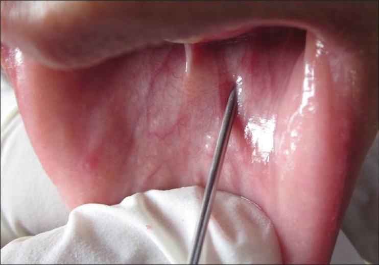
Figure 1: Oral pathergy test: mucous membrane of the lower lip is pricked using a 20 gauge blunt disposable needle - Assessment: Readings are taken after 48 h, and the test is considered positive if a pustule or ulcer is seen [Figure - 2].

Figure 2: Reading taken after 48 h shows an ulcer: positive Oral pathergy test - Sensitivity: The sensitivity of the oral PT is lower than that of the ordinary skin pathergy test.
- Advantage over the skin pathergy test: The oral PT is easier to assess than the skin PT as there is no need to measure the size of the lesion: a pustule or ulcer of any size is considered positive.
- Skin pathergy test
- Site: A hairless area on the flexor aspect of the forearms is usually chosen as the test site. [23] Ozdemir et al. [23] analyzed the skin pathergy test in different hairless body areas which included the flexor surfaces of the forearms, the lateral aspect of the tibial area, the scapular areas of the back, and the lumbar areas of the abdominal region. They concluded that the forearm is the region with the most frequently positive skin pathergy test and the abdomen the least. They suggested that variations in positivity of different body segments may result from variation in the structure, thickness and vascularity of the skin on these areas. [23]
- Various routes for skin pathergy testing: Intradermal (ID), intravenous (IV) and subcutaneous methods have been used. [24] In a study conducted by Ozlem Akmaz et al., [24] the positivity rate yielded by intradermal needles was statistically higher than that by IV application in Behcet′s patients during both the active and remission periods. [24]
- Procedure of skin pathergy test: There is no standardized pathergy test. It can be performed using 1-16 needle pricks. [23],[25],[26] In a study conducted by Ozdemir et al. [23] on the use of multiple needle pricks for skin pathergy test (SPT), they concluded that the positive pathergy reaction rates were 19%, 24%, 28%, 30%, and 33% for two, three, four, five and six needle pricks, respectively. The greatest increase in the mean percentage of positive pathergy reaction rates was detected for the application of two needle prick combination. [23] That is when two needle pricks were performed, there was a 46% increase in the positive pathergy reaction rate relative to that found with one needle prick. The percentage increase in the positive pathergy reaction rate for the three and four needle prick combinations were 26% and 17%, respectively. They concluded that two needle pricks are sufficient for SPT. [23] In most studies investigators have used either a sterile needle (20 gauge) prick or an intradermal injection of normal saline, [27] monosodium urate (MSU) crystals [28] or streptococcal antigens [7] to perform the test. The procedure is performed on a hairless part of the volar forearm. Generally, the needle is inserted vertically or diagonally at an angle of 45° to a depth of 3-5 mm. The needle should reach the dermis for a proper response. [7],[29]
- Assessment of SPT:
- Clinical evaluation: Readings are taken after 48 hrs of the needle prick. A 1-2mm papule that is usually felt by palpation and which is surrounded by an erythematous halo is formed on the skin. The papule may remain as a papule or transform into a 1-5mm pustule. The pustule becomes prominent in 24 h, becomes maximum in size in 48 h, and disappears in 45 days. Erythema without induration is interpreted as a negative result. [30]
- Histopathological evaluation: Histopa-thological studies have been performed on the skin pathergy test induced by various methods and evaluated after different time periods. Different results have been reported in various studies. They range from mononuclear cell infiltration of varying densities in perivascular and periadnexal locations with increased numbers of mast cells to leukocytoclastic vasculitis or Sweet-like vasculitis. [27] It has been suggested that the variability of the reported histopathologic features of SPT may be related to the individual differences in immune responses to inciting agents or to the different stages of skin response. [31] A more recent study compared histopathologic and clinical evaluations of the skin pathergy test and concluded that histopathological investigation was no more sensitive than clinical evaluation. [24]
- Photographic evaluation: This method was found to have increased variability and decreased sensitivity. [32]
- Factors affecting the sensitivity and specificity of skin pathergy test:
- The specificity of skin pathergy reaction is very high, especially when it is positive at 48 h, [33] although it cannot be reproduced consistently even in the same patient. Because of its specificity, the skin pathergy reaction was included in the diagnostic criteria proposed by the International Study Group for Behçet′s disease. [5] The low sensitivity of pathergy test prevents it from being used as a screening test. [34]
- Studies have demonstrated that use of a blunt (reusable, sterilized) needle increases the frequency and intensity of a positive skin pathergy test. [25] It is proposed that the reusable needles that are repeatedly sterilized in boiling water become rough due to the collection of calcium on the bent parts of the needles making them more traumatic than disposable ones. [35] However, the use of disposable needles for the prevention of diseases such as AIDS and Hepatitis B and the lesser amount of trauma initiated by these seem to decrease the rate of pathergy positivity. [35]
- The positivity rate of pathergy test in Behcet′s disease was found to vary from country to country (It is most prevalent around the silk route, which extends from the Far East to the Mediterranean Basin, including the Gulf area than in comparison to its western counterparts, especially in Europe and the USA).[24]
- Another method of increasing the rate of positivity consists of resting the needle in the dermis for 90 seconds before taking it out. [36]
- Males who were affected by Behcet′s disease were found to have a higher positivity rate than in comparison to females. [34],[37]
- Pathergy positivity is further related to the diameter of the needle. [38] A 20G disposable needle gave a positive skin pathergy test in 62.5% of cases. This figure fell to 35.8% when 26 gauge needles were used. [35] It appears that a fine needle causes insufficient trauma to induce pathergy in the dermis.
- It was shown that surgical cleaning of the skin surface before application of the needle reduced the test positivity. Some substances, bacteria, or skin products, eliminated by surgical cleaning, might play a role in the development of skin pathergy reaction.[39]
Conditions with positive pathergy phenomenon:
- Behcet′s disease [40]
- Pyoderma gangrenosum (PG): The pathergy test positivity at a rate of 25% has been reported in the literature in PG patients. [41] Aggressive surgical debridement or skin grafting is discouraged in these patients because of the risk of a pathergic response.
- Interferon alpha-treated chronic myeloid leukemia patients [42]
- Sweets syndrome [43],[44]
- Eosinophilic pustular folliculitis [45]
- Inflammatory bowel disease [46]
- Healthy individuals [47]
- Rarely in spondyloarthropathies [48]
Uses of pathergy testing:
- Positive pathergy reaction is very important for the diagnosis of Behcet′s disease in patients with only recurrent oral ulceration plus one of the other criteria (recurrent genital ulceration, eye lesions, skin lesions). [49],[50]
- The test has also been used as an indicator of disease activity in patients with Behcet′s disease. [41]
- To determine the etiological factor in a case of recurrent aphthous stomatitis. [49],[50]
- Positive pathergy test is an independent risk factor for occurrence of postoperative complications in patients with Behcet′s disease. In clinical practice, the presence of ischemia from thromboembolism, an impending vessel wall or aneurysm rupture, and bowel perforation all mandate urgent surgical intervention. In these cases, pathergy test results can be helpful to identify subgroups of patients at particular risk for postoperative complications and can guide initiation of immunosuppressive treatment in these patients. [51]
Sites of pathergy other than the skin
The pathergy phenomenon is not only restricted to the skin. In fact, any disruption of tissue integrity is potentially associated with an exaggerated inflammatory response in Behcet′s disease. In particular, the posttraumatic arterial thrombus and/or aneurysm formation following conventional angiographic interventions, [52] vascular surgery, [52],[54] superficial thrombophlebitis induced by venipuncture, eye inflammation after intraocular corticosteroid injections [55] and anastomotic ulcers following surgical treatment of intestinal ulcer [56] are well known examples of pathergy reactions triggered at different tissue sites.
Conclusion
Although the pathergy test has lost some of its sensitivity during the past 35 years, it has not lost its value as a diagnostic test. In a practical view, the chances of getting a positive test have decreased over the time. However, a positive test is rather a synonym of Behcet′s disease, with a probability of 98.4% specificity. [19]
| 1. |
Davatchi F. Behcet's disease. In: Syngle A, Deodhar SD, editors. Rheumatology principles and practice. New Delhi: Jaypee Brothers; 2009. p. 249-68.
[Google Scholar]
|
| 2. |
Dilsen N, Konice M, Aral O. Our diagnostic criteria of Behcet's disease -an overview. In: International Congress Series Lehner T, Barnes CG, editors. Recent advances in Behcet's Disease. London: Royal Society of Medicine Services;1986. p.177-80.
[Google Scholar]
|
| 3. |
Behcet's Disease Research Committee of Japan, Behcet's disease: Guide to diagnosis of Behcet's disease. Jpn J Ophthalmol 1974;18:291-4.
[Google Scholar]
|
| 4. |
Mizushima Y. Recent research into Behcet's disease in Japan. Int J Tissue React 1988;10:59-65.
[Google Scholar]
|
| 5. |
Criteria for diagnosis of Behcet's disease. International Study Group for Behcet's Disease. Lancet 1990;335:1078-80.
[Google Scholar]
|
| 6. |
Davatchi, F, Shahram, F, Akbarian M. Accuracy of existing diagnosis criteria for Behcet's Disease. In: Wechsler B, Godeau P, editors. Behcet's disease. Amsterdam: Excerpta Medica; 1993. p. 225-8.
[Google Scholar]
|
| 7. |
Davatchi F, Shahram F, Akbarian M. Classification Tree for the diagnosis of Behcet's disease. In: Wechsler B, Godeau P, editors. Behcet's disease. Amsterdam: Excerpta Medica; 1993. p. 245-8.
[Google Scholar]
|
| 8. |
Blobner F. Zur rezidivierenden Hypopyon-Iritis. Z Augenheilk 1937;91:129.
[Google Scholar]
|
| 9. |
Katzenellenbogen I, Feuerman EJ. Data on Behcet's disease (significance of the specific skin hyperreactivity and of the Behcetin reaction). Hautarzt 1965;16:13-8.
[Google Scholar]
|
| 10. |
Alekberova Z, Madanat W, Prokaeva T, Yermakova N, Poljanskaja I. Clinical and genetic features of 35 patients with Behcet's disease from commonwealth independent states. In: Wechsler B, Godeau P, editors. Behcet's disease. Amsterdam: Elsevier Science;1993. p.171-4.
[Google Scholar]
|
| 11. |
Benamour S, Zeroual B, Bennis R, Amraoui A, Bettal S. Behçet's disease. 316 cases. Presse Med 1990;19:1485-9.
[Google Scholar]
|
| 12. |
Al-Rawi ZS, Sharquie KE, Khalifa SJ, Al-Hadithi FM, Munir JJ. Behcet's disease in Iraqi patients. Ann Rheum Dis 1986;45:987-90.
[Google Scholar]
|
| 13. |
Dong Yi, Ming Qiu Xiao, Zhang Nai Zheng, Li Cui Hai,Wu Qi Yan. Testing different diagnostic criteria of Behcet's syndrome in Chinese patients. In: O'Duffy JD, Kokmen E, editors. Behcet's disease basic and clinical aspects. New York: Marcel Decker; 1991. p. 55-9.
[Google Scholar]
|
| 14. |
Assad-Khalil SH. Clinical, genetic, immunological, and biochemical features of 180 Egyptian patients with Behcet's disease. In: O'Duffy JD, Kokmen E, editors. Behcet's disease basic and clinical aspects New York: Marcel Decker Inc; 1991. p. 269-77.
[Google Scholar]
|
| 15. |
Davatchi F, Shahram F, Akbarian M, Davatchi CC, Shams H, Nadji A, et al. Behcet's disease-analysis of 3443 cases. APLAR J Rheumatol 1997;1:2-5.
[Google Scholar]
|
| 16. |
Zouboulis Ch C, Djawari D, Kirch W, Keitel W, Ochsendorf F, Orfanos CE. Adamantiades-Behcet's disease in Germany. In: Godeau P, Wechsler B, editors. Behcet's disease. Amsterdam: Elsevier Science Publishers;1993. p. 193-200.
[Google Scholar]
|
| 17. |
Nakae K, Masaki F, Hashimoto T, Inaba G, Moshizuki M, Sakane T. Recent epidemiological features of Behcet's Disease in Japan. In: Godeau P, Wechsler B, editors. Behcet's disease. Amsterdam: Elsevier Science;1993. p. 145-51.
[Google Scholar]
|
| 18. |
al-Dalaan AN, al Balaa SR, el Ramahi K, al-Kawi Z, Bohlega S, Bahabri S, et al. Behcet's disease in Saudi Arabia. J Rheumatol 1994;21:658-61.
[Google Scholar]
|
| 19. |
Davatchi F, Chams-Davatchi C, Ghodsi Z, Shahram F, Nadji A, Shams H, et al. Diagnostic value of pathergy test in Behcet's disease according to the change of incidence over the time. Clin Rheumatol 2011 [In press].
[Google Scholar]
|
| 20. |
Gül A, Esin S, Dilsen N, Koniçe M, Wigzell H, Biberfeld P. Immunohistology of skin pathergy reaction in Behcet's disease. Br J Dermatol 1995;132:901-7.
[Google Scholar]
|
| 21. |
Inaloz HS, Evereklioglu C, Unal B, Kirtak N, Eralp A, Inaloz SS. The significance of immunohistochemistry in the skin pathergy reaction of patients with Behcet's syndrome. J Eur Acad Dermatol Venereol 2004;18:56-61.
[Google Scholar]
|
| 22. |
Sharquie KE, Al-Araji A, Hatem A. Oral pathergy test in Behcet's disease. Br J Dermatol 2002;146:168-9.
[Google Scholar]
|
| 23. |
Ozdemir M, Balevi S, Deniz F, Mevlitoðlu I. Pathergy reaction in different body areas in Behcet's disease. Clin Exp Dermatol 2007;32:85-7.
[Google Scholar]
|
| 24. |
Akmaz O, Erel A, Gürer MA. Comparison of histopathologic and clinical evaluations of pathergy test in Behçet's disease. Int J Dermatol 2000;39:121-5.
[Google Scholar]
|
| 25. |
Dilºen N, Koniçe M, Aral O, Ocal L, Inanç M, Gül A. Comparative study of the skin pathergy test with blunt and sharp needles in Behcet's disease: Confirmed specificity but decreased sensitivity with sharp needles. Ann Rheum Dis 1993;52:823-5.
[Google Scholar]
|
| 26. |
Askari A, Al-Aboosi M, Sawalha A. Evaluation of pathergy test in North Jordan. Clin Rheumatol 2000;19:249-51.
[Google Scholar]
|
| 27. |
Jorizzo JL, Solomon AR, Cavallo T. Behcet's syndrome. Immunopathologic and histopathologic assessment of pathergy lesions is useful in diagnosis and follow-up. Arch Pathol Lab Med 1985;109:747-51.
[Google Scholar]
|
| 28. |
Fresko I, Ozsoy Y, Mat C, Melikoglu M, Tunc R, Yazici H. The response to the intradermal injection to monosodium urate in Behçet's syndrome and its comparison to the pathergy test. Yonsei Med J 2000;41:S25.
[Google Scholar]
|
| 29. |
Serdaroglu S, Iscimen A, Tuzun Y, Yazýcý H. The importanceof multiple needle pricks in the determination of the pathergy test in Behcet's disease. In: Memisoglu H, Acar A, editors. 13th National congress of Dermatology. vol 1. Adana: Cukurova Universities Basimevi; 1991. p. 339-44.
[Google Scholar]
|
| 30. |
Nazzaro P. Cutaneous manifestations of Behcet's disease. In: Monacelli M, Nazzaro P, editors. International symposium on Behcet's Disease in Rome. Basel: Karger; 1966. p. 1-24.
[Google Scholar]
|
| 31. |
Gilhar A, Winterstein G, Turani H, Landau J, Etzioni A. Skin hyperreactivity response (pathergy) in Behcet's disease. J Am Acad Dermatol 1989;21:547-52.
[Google Scholar]
|
| 32. |
Yazici H, Chamberlain MA, Tüzün Y, Yurdakul S, Müftüoglu A. A comparative study of the pathergy reaction among Turkish and British patients with Behcet's disease. Ann Rheum Dis 1984;43:74-5.
[Google Scholar]
|
| 33. |
Friedman-Birnbaum R, Bergman R, Aizen E. Sensitivity and specificity of pathergy test results in Israeli patients with Behcet's disease. Cutis 1990;45:261-4.
[Google Scholar]
|
| 34. |
Saylan T, Mat C, Fresko I, Melikoðlu M. Behcet's disease in the Middle East. Clin Dermatol 1999;17:209-23.
[Google Scholar]
|
| 35. |
Ozarmagan G, Saylan T, Azizlerli G, Ovül C, Aksungur VL. Re-evaluation of the pathergy test in Behcet's disease. Acta Derm Venereol 1991;71:75-6.
[Google Scholar]
|
| 36. |
Mat CM, Gokler G, Yurdakul S, Tüzün Y, Hamuryudan V, Kosem V, et al. The effect of prick duration on pathergy positivity in Behcet's syndrome. Cerrahpasa J Med 1996;27:94-7.
[Google Scholar]
|
| 37. |
Yazici H, Tüzün Y, Tanman AB, Yurdakul S, Serdaroglu S, Pazarli H, et al. Male patients with Behcet's syndrome have stronger pathergy reactions. Clin Exp Rheumatol 1985;3:137-41.
[Google Scholar]
|
| 38. |
Dilsen N, Konice M, Aral O, Aykut S. Standardization and evaluation of the skin pathergy test in Behcet's disease and controls. In: Lehner T, Barnes C, editors. Recent advances in Behcet's disease. London: Royal Society of Medicine Services; 1986. p. 177-80.
[Google Scholar]
|
| 39. |
Fresko I, Yazici H, Bayramicli M, Yurdakul S, Mat C. Effect of surgical cleaning of the skin on the pathergy phenomenon in Behcet's syndrome. Ann Rheum Dis 1993;52:619-20.
[Google Scholar]
|
| 40. |
Varol A, Seifert O, Anderson CD. The skin pathergy test: Innately useful? Arch Dermatol Res 2010;302:155-68.
[Google Scholar]
|
| 41. |
Su WP, Davis MD, Weenig RH, Powell FC, Perry HO. Pyoderma gangrenosum: Clinicopathologic correlation and proposed diagnostic criteria. Int J Dermatol 2004;43:790-800.
[Google Scholar]
|
| 42. |
Budak-Alpdogan T, Demirçay, Alpdogan O, Direskeneli H, Ergun T, Oztürk A, et al. Skin hyperreactivity of Behcet's patients (pathergy reaction) is also positive in interferon alpha-treated chronic myeloid leukaemia patients, indicating similarly altered neutrophil functions in both disorders. Br J Rheumatol 1998;37:1148-51.
[Google Scholar]
|
| 43. |
Bi XL, Gu J, Yan M, Gao CF. A case of Sweet's syndrome with slack skin and pathergy phenomenon. Int J Dermatol 2008;47:842-4.
[Google Scholar]
|
| 44. |
Awan F, Hamadani M, Devine S. Paraneoplastic Sweet's syndrome and the pathergy phenomenon. Ann Hematol 2007;86:613-4.
[Google Scholar]
|
| 45. |
Hsu PJ, Huang CJ, Wu MT. Pathergy in atypical eosinophilic pustular folliculitis. Int J Dermatol 2005;44:203-5.
[Google Scholar]
|
| 46. |
Hatemi I, Hatemi G, Celik AF, Melikoglu M, Arzuhal N, Mat C, et al. Frequency of pathergy phenomenon and other features of Behcet's syndrome among patients with inflammatory bowel disease. Clin Exp Rheumatol 2008;26:S91-5.
[Google Scholar]
|
| 47. |
Aral O, Dilsen N, Koniçe M. Positive skin pathergy reactivity as a genetic marker of Behcet's disease. In: Lehner T, Barnes C, editors. Recent advances in Behcet's disease. London: Royal Society of Medicine Services;1986. p. 173-5.
[Google Scholar]
|
| 48. |
Kaklamani V, Vaiopoulos G, Kaklamanis P. Behcet's disease. Semin Arthritis Rheum 1998;27:197-217.
[Google Scholar]
|
| 49. |
Chang HK, Kim SY. Survey and validation of the criteria for Behcet's disease recently used in Korea: A suggestion for modification of the international study group criteria. J Korean Med Sci 2003;18:88-92.
[Google Scholar]
|
| 50. |
Tunç R, Uluhan A, Melikoðlu M, Ozyazgan Y, Ozdoðan H, Yazici H. A reassesment of the international study group criteria for the diagnosis (classification) of Behcet's syndrome. Clin Exp Rheumatol 2001;19:S45-7.
[Google Scholar]
|
| 51. |
Park MC, Hong BK, Kwon HM, Hong YS. Surgical outcomes and risk factors for postoperative complications in patients with Behcet's disease. Clin Rheumatol 2007;26:1475-80.
[Google Scholar]
|
| 52. |
Alpagut U, Ugurlucan M, Dayioglu E. Major arterial involvement and review of Behcet's disease. Ann Vasc Surg 2007;21:232-9.
[Google Scholar]
|
| 53. |
Tüzün H, Sayin A, Karaözbek Y, Erdað A, Coskun H, Vural FS. Peripheral aneurysms in Behcet's disease. Cardiovasc Surg 1993;1:220-4.
[Google Scholar]
|
| 54. |
Tüzün H, Besirli K, Sayin A, Vural FS, Hamuryudan V, Hizli N, et al. Management of aneurysms in Behcet's syndrome: an analysis of 24 patients. Surgery 1997;121:150-6.
[Google Scholar]
|
| 55. |
Yalcindag FN, Batioglu F. Pathergy-like reaction following intravitreal triamcinolone acetonide injection in a patient with Behcet's disease. Ocul Immunol Inflamm 2008;16:181-3.
[Google Scholar]
|
| 56. |
Choi IJ, Kim JS, Cha SD, Jung HC, Park JG, Song IS, et al. Long-term clinical course and prognostic factors in intestinal Behcet's disease. Dis Colon Rectum 2000;43:692-700.
[Google Scholar]
|
Fulltext Views
21,068
PDF downloads
4,536


![[Figure - 1]](#fig_ijdvl_2011_77_4_526_82399_u1.jpg)
![[Figure - 2]](#fig_ijdvl_2011_77_4_526_82399_u2.jpg)