Translate this page into:
Identification of clinical and immunological factors associated with clinical relapse of pemphigus vulgaris in remission
2 Department of Endocrinology (Division of Immunology), Postgraduate Institute of Medical Education and Research, Chandigarh, India
3 Department of Histopathology, Postgraduate Institute of Medical Education and Research, Chandigarh, India
4 Department of Biostatistics, Postgraduate Institute of Medical Education and Research, Chandigarh, India
Correspondence Address:
Dipankar De
Department of Dermatology, Venereology and Leprology, Postgraduate Institute of Medical Education and Research, Sector 12, Chandigarh - 160 012
India
| How to cite this article: Guliani A, De D, Handa S, Mahajan R, Sachdeva N, Radotra BD, Kishore K. Identification of clinical and immunological factors associated with clinical relapse of pemphigus vulgaris in remission. Indian J Dermatol Venereol Leprol 2020;86:233-239 |
Abstract
Background: Pemphigus vulgaris is a potentially fatal autoimmune epidermal blistering disease with a chronic and relapsing course. It is difficult to predict clinical relapse. Identification of clinical and immunological factors that are associated with early clinical relapse in a prospective study design may help in planning treatment for better maintenance of clinical remission.
Aim: The aim of our study was to identify clinical and immunological factors associated with clinical relapse within 9 months of study inclusion in patients with pemphigus vulgaris in clinical remission.
Methods: Forty consecutive consenting patients who had been diagnosed to have pemphigus vulgaris and were in clinical remission on minimal therapy or off therapy were included. The patients were followed up every 3 months until 9 months. Clinical factors considered relevant were recorded at the beginning of the study. Immunological factors such as CD19+ B-cell count and CD19+CD27+ memory B cells/plasma cell count in peripheral blood were assessed at baseline [anti-desmoglein (Dsg) 1 and 3 titers were first assessed at 3 months, not at baseline] and repeated every 3 months, until 9 months or clinical relapse whichever was earlier. Direct immunofluorescence (DIF) of skin biopsy specimen was performed at study initiation and again at the time of clinical relapse or study completion, whichever occurred earlier. All patients completed the study.
Results: Of 40 patients, 11 (27.5%) experienced relapse as per definition, while 29 (72.5%) remained in complete remission. Clinical relapse during study duration was significantly more common in those who had onset of disease in oral mucosa [odds ratio (OR), 10.71; 95% confidence interval (CI) 1.21–94.86, P = 0.02], pruritus (OR 8.4; 95% CI 1.76–40.02, P = 0.01), and extensive cutaneous involvement during previous disease activity (OR 7.36; 95% CI 1.34–40.55, P = 0.03) and also pruritus during remission (P = 0.004). Immunological factors found to be significantly associated with early clinical relapse were raised CD19+ B-cell count at baseline (OR 7.84; 95% CI 1.39 – 53.41, P = 0.01), immunoglobulin G (OR 4.85; 95% CI 1.09–23.44, P = 0.04), and C3 (OR 20.33; 95% CI 3.02–199.5, P < 0.001) positivity in the intercellular space of the epidermis on DIF at study onset and rising anti-Dsg 3 antibody titers (OR 19.96; 95% CI 1.85- 310.9, P = 0.03).
Limitations: Limited sample size, short follow-up duration, and inability to perform anti-Dsg enzyme linked immunosorbent assay for all the patients at all the time points of assessment are limitations of this study.
Conclusion: Immunological relapse can be determined before clinical relapse, so that treatment can be restarted/modified and clinical remission can be maintained.
Introduction
Pemphigus vulgaris is an autoimmune disease marked by chronic and relapsing course. Often, low-dose immunosuppression with prednisolone (<10 mg/day) alone or in combination with immunosuppressants such as cyclophosphamide and azathioprine (<1 mg/kg/day) is continued after achieving clinical remission to maintain it and prevent relapse. This puts the patients at an increased risk of side effects.
Earlier studies have identified clinical/epidemiological factors such as older age of onset,[1],[2] number of blisters per day before starting initial treatment, duration of clinical remission before inclusion into the study,[3] oral mucosal involvement as initial presentation,[2],[4],[5] and pruritus[6] to be important determinants of disease course. Some of these studies concluded that a negative skin biopsy direct immunofluorescence (DIF) and a negative anti-desmoglein (Dsg) antibody titer by enzyme linked immunosorbent assay (ELISA) may help identify a patient in whom the treatment can be stopped. Earlier studies that have tried to identify immunological predictors of relapse had several limitations, that is, retrospective study design, only one-time immunological investigation in prospective studies, and dependence only on DIF. A recent retrospective study has studied biomarkers for prediction of clinical relapse in patients treated only with rituximab.[7]
The objective of our study was to identify clinical and immunological factors associated with early clinical relapse, arbitrarily defined as relapse within 9 months of study inclusion. In a prospective study design, we assessed clinical parameters at study initiation and periodically assessed immunological parameters in patients with pemphigus vulgaris in remission irrespective of treatment they had received. We also determined which of these parameters could be associated clinical relapse and the time interval between immunological occurrence and clinical relapse.
Methods
This prospective study was undertaken from January 2017 to October 2017 at the immunobullous disease clinic of the Postgraduate Institute of Medical Education and Research, Chandigarh, India. Patients who were in complete clinical remission defined as being lesion-free for at least 2 months while on no treatment (complete remission off treatment) or minimal treatment [defined as less than or equal to 10 mg/day of prednisone (or the equivalent) and/or minimal adjuvant therapy for at least 2 months, complete remission on minimal treatment] were included.[8] Inability to come for quarterly follow-up visits for at least 9 months, pregnant and lactating women, and elderly patients age 70 years or older were the excluding criteria in our study. At study onset, history as per clinic records or patient's personal medical records were extracted in the predesigned case record form of the study. These clinical characteristics included age at disease onset, oral mucosal involvement as initial presentation, history of pruritus anytime during disease activity, interval between disease onset and treatment initiation (current episode), total number of previous relapses, extensive cutaneous involvement [defined as> 5% body surface area (BSA) involvement], cumulative duration of treatment before achievement of clinical remission (current episode), duration of clinical remission before inclusion in the study, and pruritus during study. Immunological investigations inclusive of skin biopsy DIF, anti-Dsg ELISAs, and estimation of count in peripheral blood of CD19+ B cells and CD19+CD27+ memory B cells/plasma cells were carried out. For flow cytometric assessment of B cells, the lymphocytes were stained with fluorescein isothiocyanate–labeled anti-human CD 19 and phycoerythrin-labeled anti-human CD 27 antibodies (BD Biosciences, San Jose, CA, USA) as per manufacturer's protocol. ELISA for Dsg 1 and 3 was carried out using commercially available research-only kits (human Dsg-1 and 3 ELISA kit; Sunred Bio, Shanghai, China) as per manufacturer's protocol.
One-time assessment of CD19+ B cells and CD19+CD27+ memory B cells' count was performed in equal number of controls to have data regarding their counts in normal north Indian population.
CD 19+ B cells and CD19+CD27+ memory B cells were carried out every 3 months until relapse or study completion, whichever was earlier; skin biopsy DIF was carried out at baseline and at the end of the study or relapse, whichever was earlier; ELISA for anti-Dsg 1 and 3 antibodies was first carried out at 3 months and then every 3 months until relapse or end of the study, whichever was earlier. Clinical relapse was defined as appearance of a new lesion that did not subside spontaneously within 1 week of appearance.[8]
The study protocol was reviewed and approved by the Institute Ethics Committee (intramural) of the Postgraduate Institute of Medical Education and Research, Chandigarh, India. Informed and written consent was obtained from all patients before their inclusion in the study.
For statistical analysis, IBM SPSS 22.0 (USA) software was used. Univariate analysis for comparison of quantitative outcomes such as immunoglobulin levels and memory B-cell counts between two groups (subjects with and without relapse) was carried out using Mann–Whitney U-test. Furthermore, associations between categorical risk factors such as DIF results between subjects with and without relapse were calculated using Fisher's exact test. Odds ratio (OR) for the same was calculated using SPSS and OpenEpi (http://www.openepi.com/Menu/OE_Menu.htm) software. A nondirectional P value < 0.05 was used to declare statistical significance for all the tests in the analysis.
Results
A convenient sample of 40 patients in complete remission (off therapy or on minimal therapy) who satisfied inclusion criteria were included in the study consecutively after informed consent. Of the 40 patients, 26 were in complete remission off therapy and 14 were in complete remission on minimal therapy at the time of study initiation. For those with complete remission on minimal therapy at inclusion, the treatment was gradually tapered and finally stopped. If there was a relapse, they reached the endpoint of the study. Of the 26 patients in complete remission off therapy, 9 (34.6%) relapsed; while among 14 patients who were in complete remission on minimal therapy, 2 (14.2%) relapsed within the study duration.
The mean interval between achievement of clinical remission and inclusion in this study was 10.12 months. Of 40 participants, 11 (27.5%) had relapse during the study as per definition, while 29 (72.5%) remained in remission. Clinicodemographic characteristics, that is, mean age at disease onset, gender distribution, type of pemphigus vulgaris (mucosal or mucocutaneous), and treatment modality used to induce remission, were comparable between those who relapsed within 9 months and those who maintained remission [Table - 1].

Treatment that had been used before induction of remission is detailed in [Table - 1]. No particular treatment before achieving remission was found to be particularly associated with early relapse. None of the 11 patients who had disease relapse was on any treatment at the time of relapse.
Clinical characteristics at study onset were recorded from previous medical records. Factors not found significantly associated with relapse were age at onset, interval between disease onset and initiation of treatment, total number of previous relapses, duration of treatment before achievement of this episode of clinical remission, and duration of clinical remission before inclusion into the study. Clinical characteristics of patients are illustrated in [Table - 2].

Oral mucosa was the first site to be involved in 24 (60%) patients. Clinical relapse occurred in 10 (43.4%) out of those 24 patients who had disease onset in the oral mucosa, while 1 (11.7%) of 16 patients with onset on the skin had relapse. Oral mucosal involvement as initial manifestation was significantly more frequent among relapse group when compared with remission group [OR 10.71; 95% confidence interval (CI) 1.21–94.86, P = 0.02]. Pruritus anytime during disease activity was present in 12 (30%) patients. Clinical relapse occurred in 7 (58.33%) of those 12 patients who had pruritus, while 4 (14.28%) of 28 patients who had not have pruritus relapsed. Twenty patients each had limited (≤5% BSA) and extensive (>5% BSA) disease (at the time of most severe disease) in the preceding activity episode. Of 20 patients with> 5% BSA involvement, 9 (45%) had disease relapse; while of 20 patients with ≤5% BSA involvement, only 2 (10%) relapsed. The proportion of patients with extensive cutaneous involvement in the preceding activity episode was found to be higher in the relapse group when compared with the remission group (OR 7.36; 95% CI 1.34–40.55, P = 0.03). Pruritus anytime during disease activity was significantly common in those who had relapse in comparison to those who maintained remission (OR 8.4; 95% CI 1.76–40.02, P = 0.01). Pruritus within the duration of this study was recorded in 4 (10%) patients. All 4 (100%) patients with pruritus relapsed, while 7 (19.4%) of 36 who had no pruritus relapsed (P = 0.004).
The mean CD19 + B-cell count in controls was 4.6% ± 1.9%. Since normal value for CD19+ B cell count in north Indian population is not known, CD19+ B-cell count upto 4.6% was considered normal for analysis of results of this study, whereas more than this was considered as raised CD19+ B-cell count. The mean CD19+CD27+ memory B-cell count in controls was 1.5% ± 0.96%. Similarly, CD19+CD27+ memory B-cell count upto 1.5% was considered normal for this study, whereas more than this was considered as raised CD19+CD27+ B-cell count.
Flow cytometry data at study initiation were available for all patients. Raised CD19 + B-cell count at baseline was associated with increased risk of relapse between 0–6 and 0–9 months (OR 7.84; 95% CI 1.39–53.41, P = 0.01) as illustrated in [Table - 3]. The probability of clinical relapse in patients with raised CD19+ B-cell counts at baseline was 33.3% at 3 months increasing to 58.3% at 6 and 9 months (no patient relapsed after 6 months of the study). The CD19+CD27+ memory B-cell counts at baseline, 3 months, and 6 months were not found to be associated with relapse between 0–3 months, 3–6 months, and 6–9 months, respectively.

DIF data at the beginning of the study were available for all patients. Positive immunoglobulin G (IgG) (OR 4.85 95% CI 1.09–23.44, P = 0.04) and C3 (OR 20.33; 95% CI 3.02–199.5, P < 0.001) in the intercellular spaces of the epidermis on DIF at baseline were found to be significantly associated with clinical relapse within the study duration as illustrated in [Table - 4]. Only 13.6% of patients with negative DIF at study onset had clinical relapse in 9 months.

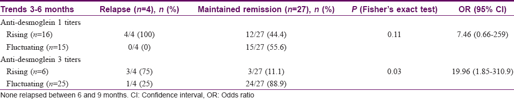
The cut-off values for assigning a positive anti-Dsg 1 or 3 antibodies titers were not provided in the research-only ELISA kits used. Hence, trends of anti-Dsg 1 and 3 antibody titers were assessed for correlation with clinical relapse. Rising trends of anti-Dsg 1 antibody titer were seen in all the patients with clinical relapse and 44.4% of those who did not relapse (OR 7.46; 95% CI 0.66–259, P = 0.11). Rising trends of anti-Dsg 3 antibody titer were found in 75% and 11.1% of patients with relapse and without relapse, respectively (OR 19.96; 95% CI 1.85-310.9, P = 0.03). Immunologic parameters in patients with relapse and maintained remission are illustrated in [Table - 5].
Assessment of time interval between immunological occurrence and clinical relapse
Among 11 patients whose disease relapsed clinically, 9 had preceding immunological occurrence in the form of raised CD19+ B cells, 6 patients at the beginning of the study, and 3 patients at 3 months. The mean time interval between completion of previous treatment and immunological event in the form of raised CD19+ B-cell count was 14.66 months. The mean interval between this immunological occurrence and clinical relapse was 2.22 ± 1.85 months.
Of 11 patients with relapse, 8 had positive DIF (thus, immunological occurrence) at study initiation and 3 had negative DIF. The interval between this immunological occurrence in these 8 patients and clinical relapses was 3.75 ± 1.66 months.
Discussion
Earlier studies that have tried to identify immunological predictors of relapse were limited by retrospective study design, only one-time immunological investigation in prospective studies, and dependence only on DIF. No prospective study has periodically followed up patients by testing immunological parameters to predict when a patient is about to have clinical relapse. The principal findings of our prospective study are oral mucosal involvement as initial presentation, pruritus anytime during disease activity, extensive cutaneous involvement during previous activity episode, and pruritus during this study are associated with early clinical relapse. Immunological factors found to be significantly associated with clinical relapse are higher CD19+ B-cell count at baseline, IgG and C3 positivity at the intercellular spaces of the epidermis on DIF at baseline, and rising anti-Dsg 3 antibody titers.
Kanwar et al. observed that pruritus may act as a clinical sign of disease activity.[6] Our study suggests that new-onset pruritus in a pemphigus patient in remission may be an indicator of disease activity and thus impending relapse. The mean interval between new onset of pruritus within the study duration and clinical relapse was 13.5 days. Extensive cutaneous involvement (>5% maximum BSA involvement) has been reported to be associated with poor prognosis and thus mortality.[2],[9] Our study suggests that severe disease may be associated with an early clinical relapse of disease.
In recent retrospective study by Albers et al., it was observed that relapse is associated with B-cell repopulation (raised CD19+ B-cell counts).[7] The investigators arbitrarily fixed the cut-off value for normal CD19+ B-cell counts instead of determining normative CD19+ B-cell counts in normal controls. We considered determination of cut-off value for normal CD19+ B-cell necessary as normal CD19+ B-cell count may vary with ethnicity. We observed that increased CD19+ B-cell count at study onset was associated with increased risk of relapse between 0–6 and 0–9 months.
In the study by Albers et al., the mean time for B cells to repopulate after treatment was 8.58 months and the mean time from repopulation to relapse was 7.99 months, with a total disease-free interval of 16.57 months.[7] Our results are somewhat similar. The mean interval between previous treatment and immunological occurrence in the form of raised CD19+ B-cell count was 14.66 months. The mean interval between the occurrence of immunological event and relapse was 2.22 months. Thus, the mean disease-free interval was 16.88 months. Albers et al. studied patients treated with rituximab only, whereas our patients were treated with rituximab or other treatment modalities. Although the results may not be directly comparable between the studies, it can be assumed that B-cell repopulation must occur irrespective of treatment modality used preceding clinical relapse in a prototype IgG-mediated disease.
Positive DIF for C3 has been reported to be associated with increased risk of clinical relapse.[2] It is suggested that negative DIF is a possible marker of cure of the disease.[10] Balighi et al. observed that patients in whom treatment was stopped 6–12 months after a negative DIF report had prolonged remissions in comparison to those in whom treatment was stopped within 6 months of a negative DIF report.[3] In our study, positive IgG and C3 at the intercellular spaces of the epidermis on DIF at study onset were found to be significantly associated with increased incidence of clinical relapse within the 9 months of study. At the time of relapse, 10 of 11 relapsing patients had positive DIF.
Rising trend of anti-Dsg 3 antibody titer was found in 75% of patients with relapse in comparison to 11.1% in patients without relapse. In a previous study, positive anti-Dsg 3 ELISA was found in patients with pemphigus vulgaris in clinical remission who relapsed earlier.[11] Either positive value or rising trend of anti-Dsg 3 when assessed periodically may act as a predictor of clinical relapse.
Limitations
Limited sample size, short follow-up duration, and inability to perform anti-Dsg ELISA for all the patients at all the time points of assessment are limitations of this study. Patients above 70 years of age were excluded because pemphigus patients are younger in India,[12] the average life expectancy at birth in India is 67.9 years,[13] and elderly in India do not have robust social support system. Follow-up was limited to a maximum of 9 months as ensuring periodic hospital visit while being under clinical remission is often difficult. Funding constraints did not allow anti-Dsg ELISA assay at all time points of assessment.
Nevertheless, in an uncommon disease such as pemphigus, 40 patients in remission following up for a planned 9 months and providing valuable insight to clinical and immunological predictors of clinical relapse in a prospectively carried out study are strengths of the study.
Conclusion
Oral mucosal involvement as initial presentation, pruritus anytime during disease activity, extensive cutaneous involvement during previous activity episode, and pruritus during this study are associated with early clinical relapse. Immunological factors found to be significantly associated with clinical relapse are higher CD19 + B-cell count at study onset, IgG and C3 positivity at the intercellular spaces of the epidermis on DIF at study onset, and rising anti-Dsg 3 antibody titers. Presence or new occurrence of these parameters can predict clinical relapse, and thus treatment can be restarted or modified resulting in better maintenance of remission in this potentially life-threatening condition.
Declaration of patient consent
The authors certify that they have obtained all appropriate patient consents. In the form the patients have given their consent for their images and other clinical information to be reported in the journal. The patients understand that their names and initials will not be published and due efforts will be made to conceal their identity, but anonymity cannot be guaranteed.
Financial support and sponsorship
'Special Research Grant' for MD thesis from PGIMER, Chandigarh.
The study protocol was approved by the Institutional Ethics Committee (Intramural) of the Postgraduate Institute of Medical Education and Research, Chandigarh.
Conflicts of interest
There are no conflicts of interest.
| 1. |
Savin JA. Some factors affecting prognosis in pemphigus vulgaris and pemphigoid. Br J Dermatol 1981;104:415-20.
[Google Scholar]
|
| 2. |
Ratnam KV, Pang BK. Pemphigus in remission: Value of negative direct immunofluorescence in management. J Am Acad Dermatol 1994;30:547-50.
[Google Scholar]
|
| 3. |
Balighi K, Taheri A, Mansoori P, Chams C. Value of direct immunofluorescence in predicting remission in pemphigus vulgaris. Int J Dermatol 2006;45:1308-11.
[Google Scholar]
|
| 4. |
Bernard P, Reguiai Z, Tancrède-Bohin E, Cordel N, Plantin P, Pauwels C, et al. Risk factors for relapse in patients with bullous pemphigoid in clinical remission: A multicenter, prospective, cohort study. Arch Dermatol 2009;145:537-42.
[Google Scholar]
|
| 5. |
Seidenbaum M, David M, Sandbank M. The course and prognosis of pemphigus. A review of 115 patients. Int J Dermatol 1988;27:580-4.
[Google Scholar]
|
| 6. |
Kanwar AJ, Gupta R, Kaur S. Pruritus – A clinical sign of activity of pemphigus. Indian J Dermatol Venereol Leprol 1989;55:396.
[Google Scholar]
|
| 7. |
Albers LN, Liu Y, Bo N, Swerlick RA, Feldman RJ. Developing biomarkers for predicting clinical relapse in pemphigus patients treated with rituximab. J Am Acad Dermatol 2017;77:1074-82.
[Google Scholar]
|
| 8. |
Murrell DF, Dick S, Ahmed AR, Amagai M, Barnadas MA, Borradori L, et al. Consensus statement on definitions of disease, end points, and therapeutic response for pemphigus. J Am Acad Dermatol 2008;58:1043-6.
[Google Scholar]
|
| 9. |
Ryan JG. Pemphigus. A 20-year survey of experience with 70 cases. Arch Dermatol 1971;104:14-20.
[Google Scholar]
|
| 10. |
David M, Weissman-Katzenelson V, Ben-Chetrit A, Hazaz B, Ingber A, Sandbank M. The usefulness of immunofluorescent tests in pemphigus patients in clinical remission. Br J Dermatol 1989;120:391-5.
[Google Scholar]
|
| 11. |
Daneshpazhooh M, Zafarmand Sedigh V, Balighi K, Hosseini SH, Ramezani A, Kalantari MS, et al. Immunologic prediction of relapse in patients with pemphigus vulgaris (PV) in clinical remission. J Am Acad Dermatol 2016;74:1160-5.
[Google Scholar]
|
| 12. |
Kanwar AJ, De D. Pemphigus in India. Indian J Dermatol Venereol Leprol 2011;77:439-49.
[Google Scholar]
|
| 13. |
Available from: https://niti.gov.in/content/life-expectancy. (last accessed on July 9, 2019)
[Google Scholar]
|
Fulltext Views
4,125
PDF downloads
1,449


![[Table - 1]](#tbl_ijdvl_2020_86_3_233_273926_t1.jpg)
![[Table - 2]](#tbl_ijdvl_2020_86_3_233_273926_t2.jpg)
![[Table - 3]](#tbl_ijdvl_2020_86_3_233_273926_t3.jpg)
![[Table - 4]](#tbl_ijdvl_2020_86_3_233_273926_t4.jpg)
![[Table - 5]](#tbl_ijdvl_2020_86_3_233_273926_t5.jpg)