Translate this page into:
Dermatofibrosarcoma protuberans: Two rare variants
2 Dr Dashore's DHL Clinic, Indore, Madhya Pradesh, India
3 Singhania Skin Clinic, Raipur, Chhattisgarh, India
Correspondence Address:
Sarina Jain
Department of Dermatology, Main Hospital Building, Ward 17-18, Ground Floor, KEM Hospital, Parel, Mumbai - 400 012, Maharashtra
India
| How to cite this article: Jain S, Dashore S, Singhania B, Khopkar US, Darkase B. Dermatofibrosarcoma protuberans: Two rare variants. Indian J Dermatol Venereol Leprol 2019;85:204-208 |
Sir,
Dermatofibrosarcoma protuberans is a dermal neoplasm with low malignant potential. Histopathologically, it is a poorly circumscribed neoplasm characterized by storiform arrangement of spindle-shaped cells in short interlacing fascicles.[1] We hereby describe two rare variants of dermatofibrosarcoma protuberans.
Case 1
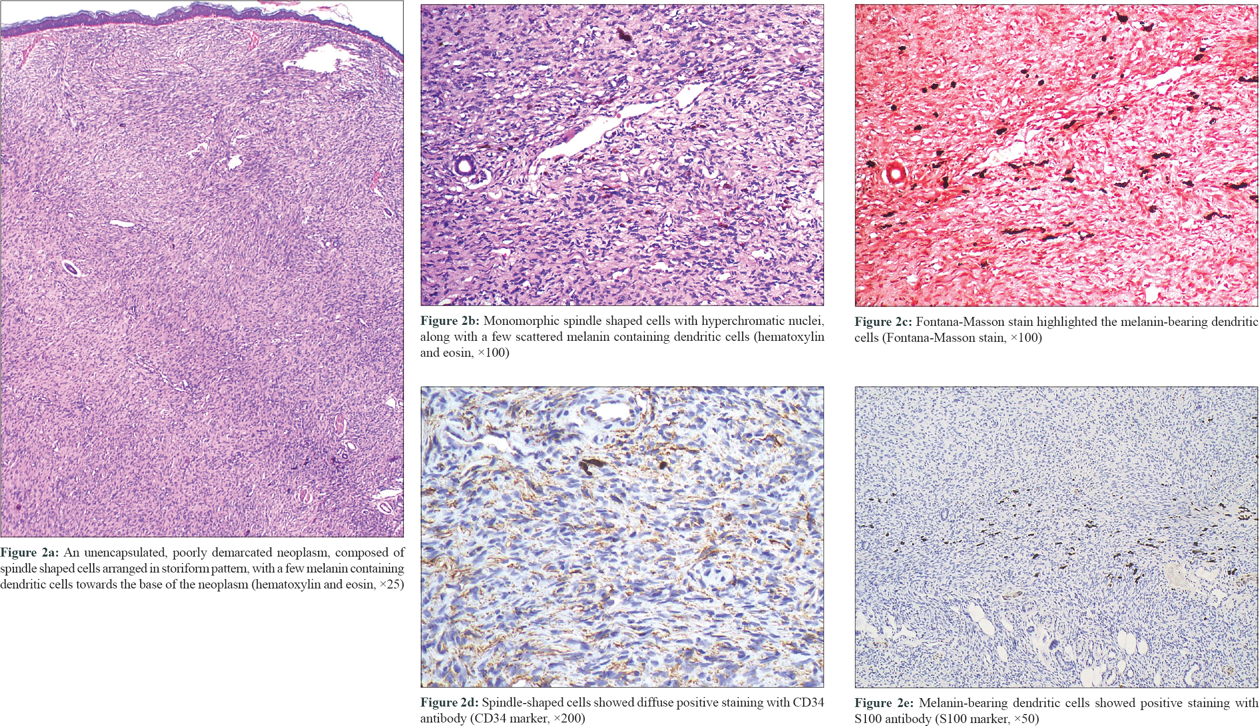
A 42-year-old woman presented with an asymptomatic, slowly progressive lesion on her right shoulder since last 25 years. Dermatological examination revealed a single ill-defined, atrophic, indurated, nontender, erythematous papulonodular plaque sized 6 cm × 4 cm with few intervening areas of bluish discoloration [Figure - 1]. Punch biopsy from an erythematous nodule demonstrated an unencapsulated, poorly demarcated neoplasm extending from the papillary dermis to the base of specimen, thus obscuring the entire dermis and subcutis. [Figure - 2]a. The overlying epidermis showed flattening and loss of rete ridges. The neoplasm was densely packed with spindle-shaped cells arranged in multiple, irregular, interwoven fascicles, resulting in a storiform pattern. These spindle-shaped cells were monomorphic their hyperchromatic slender nuclei showing negligible pleomorphism [Figure - 2]b. Near the lower end, some interspersed melanin-containing dendritic cells were noted after appropriate staining (Fontana-Masson stain). [Figure - 2]c. Immunohistochemical analysis confirmed the presence of two distinct cell populations- the spindle-shaped cells showing diffuse positive staining with CD34 and negative staining with S100 [Figure - 2]d while the melanin-bearing cells stained positively with S100 and negative for CD34 [Figure - 2]e.
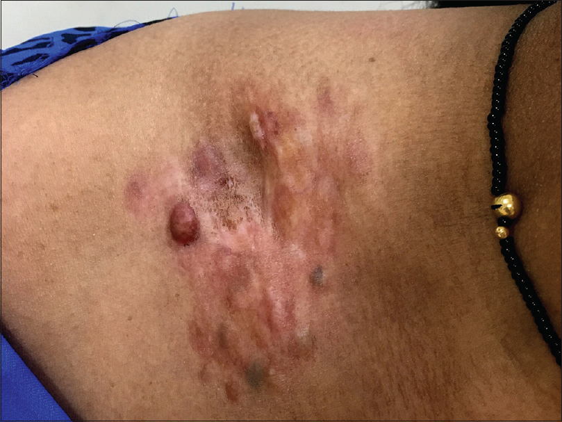 |
| Figure 1: An ill-defined, atrophic, indurated, erythematous papulonodular plaque with some areas of bluish discoloration present on the right shoulder |
 |
Based on the above findings, a diagnosis of Bednar's tumor (pigmented dermatofibrosarcoma protuberans) was made.
Case 2
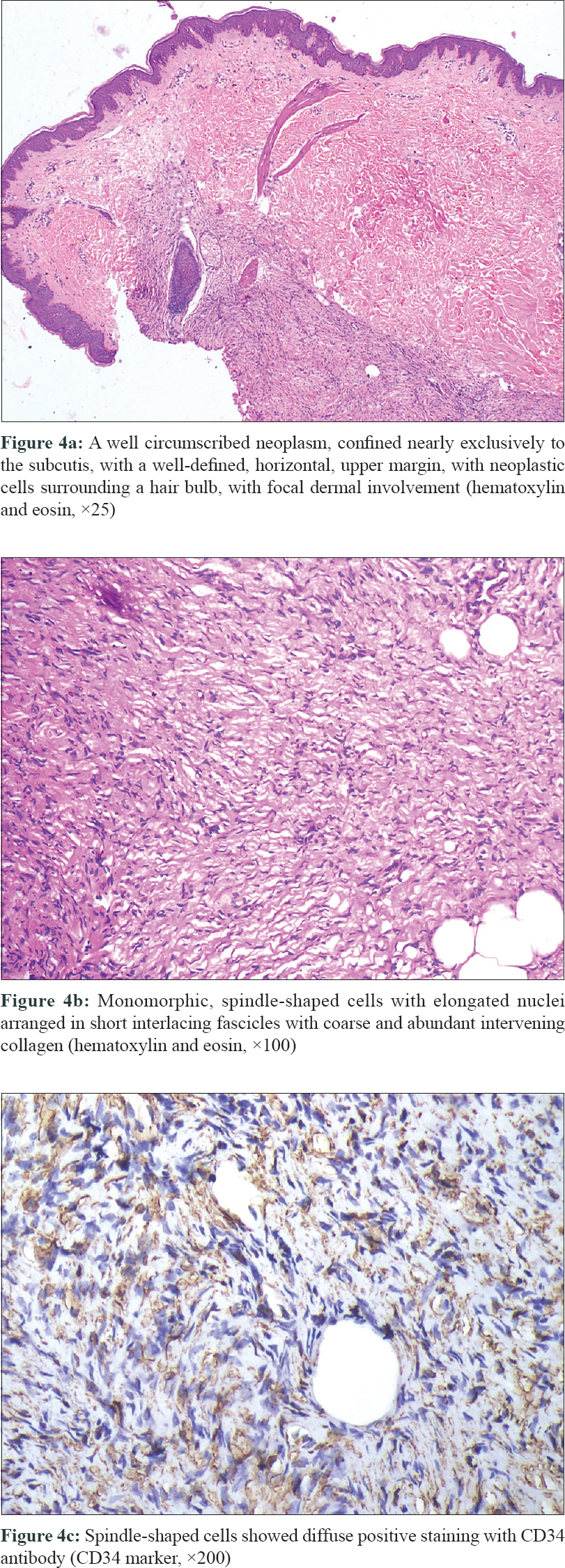
A 19-year-old woman presented with an asymptomatic lump on her right thigh for the last 5 years gradually increasing in size. On local inspection, the overlying skin was normal without any visible lump. However, palpation revealed a deep, firm, ill-defined, nontender, movable subcutaneous mass with proportions 3 cm × 5 cm [Figure - 3]. Morphea and subcutaneous zygomycosis were considered as differentials. Histopathological examination revealed a well-circumscribed neoplasm, though microscopically infiltrative, limited almost exclusively to the subcutis, with a well-defined horizontal, upper margin [Figure - 4]a. The neoplasm was composed of monomorphic, spindle-shaped cells with elongated non-pleomorphic nuclei, arranged in short interlacing fascicles [Figure - 4]b. The intervening collagen was coarse and abundant. The neoplasm infiltrated the subcutis in a honey-comb pattern reaching the base of the specimen. The neoplastic cells also surrounded a hair bulb with focal dermal involvement [Figure - 4]a. On immunohistochemical analysis, the neoplastic cells stained diffusely positive with CD34 and negative with S100 [Figure - 4]c.
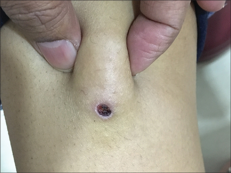 |
| Figure 3: Palpation revealed a deep, firm, ill-defined subcutaneous mass on the right thigh |
 |
On the basis of above analysis, a diagnosis of subcutaneous dermatofibrosarcoma protuberans was made. Bednar's tumor (pigmented dermatofibrosarcoma protuberans) represents 1–5% of all dermatofibrosarcoma protuberans cases.[2] It differs from classic dermatofibrosarcoma protuberans only by the presence of scattered melanin-bearing dendritic cells. Clinically, t can be nonpigmented or pigmented (blue/black), depending on the amount of melanin pigment.[1] The histogenesis of melanin-containing dendritic cells remain controversial. Neuroectodermal differentiation and melanocytic colonization from epidermis are the two proposed theories.[3], Neuromesenchymal origin has also been suggested, as corroborated by the association of Bednar's tumor with dermal melanocytosis (nevus of Ito).[4]
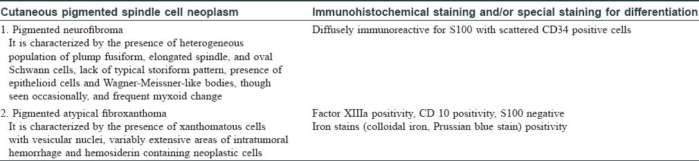
Histopathologically, it is characterized by spindle-shaped cells, arranged in a storiform pattern, admixed with a variable population of melanin-containing dendritic cells. On immunohistochemistry, spindle-shaped cells and the melanin-containing dendritic cells stain positively with CD34 and S100 respectively. Histopathological differential diagnoses include other pigmented spindle cell neoplasms [Table - 1].
Conventionally, dermatofibrosarcoma protuberans is a dermal neoplasm with frequent extension into subcutaneous fat; however, recently, a new variant, subcutaneous dermatofibrosarcoma protuberans has been recognized, where the neoplasm is confined exclusively or almost exclusively to subcutaneous fat with minimal and clinically inapparent dermal involvement.
Clinically, subcutaneous dermatofibrosarcoma protuberans manifests as an asymptomatic, nonprotuberant, nonatrophic, well- or ill-defined, indurated, subcutaneous mass without any visible skin change.[5] Histopathologically, it is characterized by a dense storiform arrangement of monomorphic spindle-shaped cells with slender nuclei, confined almost exclusively to the subcutis. Another unusual feature of this variant is its frequently well-circumscribed nature (although microscopically infiltrative). Bague and Folpe documented 15 cases of subcutaneous dermatofibrosarcoma protuberans, and found all the cases to be well-circumscribed.[6] Recently, Llombart et al. studied 18 cases of subcutaneous dermatofibrosarcoma protuberans and observed 10 cases to be well circumscribed and eight cases as poorly delimited and infiltrative.[5]
Immunohistochemistry with CD34 antibody reveals the neoplastic cells to be diffusely positive.[5],[6] Hence, except for the deep location and well-circumscribed nature, the histopathological and immunohistochemical features of subcutaneous dermatofibrosarcoma protuberans are identical to cutaneous (conventional) dermatofibrosarcoma protuberans.
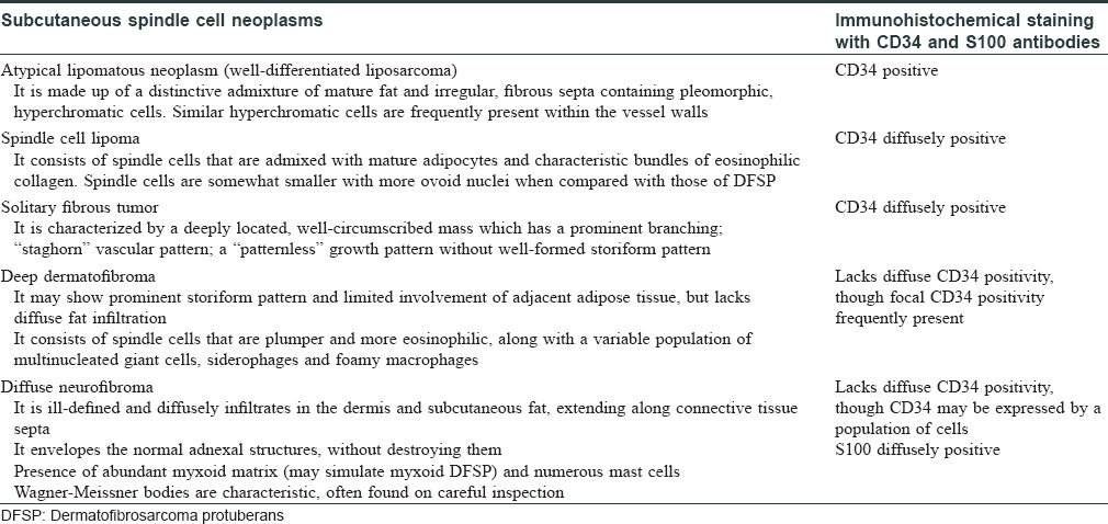
Histopathological differential diagnoses of subcutaneous dermatofibrosarcoma protuberans include other subcutaneous spindle cell neoplasms [Table - 2].[6] These cases highlight the importance of careful recognition of stereotypical histopathological features of dermatofibrosarcoma protuberans and prudent use of ancillary immunohistochemistry (CD34 antibody) to accurately diagnose these rare, unusual variants of a low malignant neoplasm. In addition, awareness of atypical clinical presentation and deep biopsy are needed to diagnose it's rarer variants.
Declaration of patient consent
The authors certify that they have obtained all appropriate patient consent forms. In the form, the patients have given their consent for their images and other clinical information to be reported in the journal. The patients understand that name and initials will not be published and due efforts will be made to conceal identity, but anonymity cannot be guaranteed.
Financial support and sponsorship
Nil.
Conflicts of interest
There are no conflicts of interest.
| 1. |
Llombart B, Serra-Guillén C, Monteagudo C, López Guerrero JA, Sanmartín O. Dermatofibrosarcoma protuberans: A comprehensive review and update on diagnosis and management. Semin Diagn Pathol 2013;30:13-28.
[Google Scholar]
|
| 2. |
Dupree WB, Langloss JM, Weiss SW. Pigmented dermatofibrosarcoma protuberans (Bednar tumor): A pathologic, ultrastructural, and immunohistochemical study. Am J Surg Pathol 1985;9:630-9.
[Google Scholar]
|
| 3. |
Ding JA, Hashimoto H, Sugimoto T, Tsuneyoshi M, Enjoji M. Bednar tumor (pigmented dermatofibrosarcoma protuberans). An analysis of six cases. Acta Pathol Jpn 1990;40:744-54.
[Google Scholar]
|
| 4. |
Amonkar GP, Rupani A, Shah A, Deshpande R. Bednar tumor: An uncommon entity. Dermatopathology (Basel) 2016;3:36-8.
[Google Scholar]
|
| 5. |
Llombart B, Serra-Guillén C, Rubio L, Nagore E, Requena C, Traves V, et al. Subcutaneous dermatofibrosarcoma protuberans, a rare subtype with predilection for the head: A retrospective series of 18 cases. J Am Acad Dermatol 2017;77:503-110.
[Google Scholar]
|
| 6. |
Bague S, Folpe AL. Dermatofibrosarcoma protuberans presenting as a subcutaneous mass: A clinicopathological study of 15 cases with exclusive or near-exclusive subcutaneous involvement. Am J Dermatopathol 2008;30:327-32.
[Google Scholar]
|
Fulltext Views
3,576
PDF downloads
1,123


![[Figure - 1]](#fig_ijdvl_2019_85_2_204_251208_f1.jpg)
![[Figure - 2]](#fig_ijdvl_2019_85_2_204_251208_f2.jpg)
![[Figure - 3]](#fig_ijdvl_2019_85_2_204_251208_f3.jpg)
![[Figure - 4]](#fig_ijdvl_2019_85_2_204_251208_f4.jpg)
![[Table - 1]](#tbl_ijdvl_2019_85_2_204_251208_t5.jpg)
![[Table - 2]](#tbl_ijdvl_2019_85_2_204_251208_t6.jpg)