Translate this page into:
Dermatoscopic evaluation of three cases of nevus lipomatosus cutaneous superficialis
2 Department of Histopathology, Postgraduate Institute of Medical Education and Research, Chandigarh, India
Corresponding Author:
Muthu Sendhil Kumaran
Department of Dermatology, Venereology and Leprology, Postgraduate Institute of Medical Education and Research, Sector 12, Chandigarh - 160 012
India
drsen_2000@yahoo.com
| How to cite this article: Vinay K, Sawatkar GU, Saikia UN, Kumaran MS. Dermatoscopic evaluation of three cases of nevus lipomatosus cutaneous superficialis. Indian J Dermatol Venereol Leprol 2017;83:383-386 |
Sir,
Nevus lipomatosus cutaneous superficialis is an uncommon hamartoma characterized by ectopic presence of mature adipocytes in the papillary dermis. The use of dermatoscopy has extended beyond skin cancers and is increasingly being utilized as a diagnostic aid in many benign and inflammatory conditions.[1] We herein describe the dermatoscopic features of three cases of nevus lipomatosus cutaneous superficialis. We also report an index case on an unusual site in which dermatoscopy aided in establishing the diagnosis.
A 20-year-old lady presented to the dermatology outpatient department with an asymptomatic lesion on the nape of her neck which was gradually progressive over the past 7 years. Cutaneous examination revealed a skin colored, well-defined plaque of 30 cm × 15 cm size extending from the lower scalp hair margin to the nape of the neck on the left side [Figure 1a]. It had an irregular surface and was studded with multiple comedones [Figure 1a]. A punch skin biopsy showed globules of mature adipocytes deposited between the normal collagen fibers of the papillary dermis [Figure 1b].
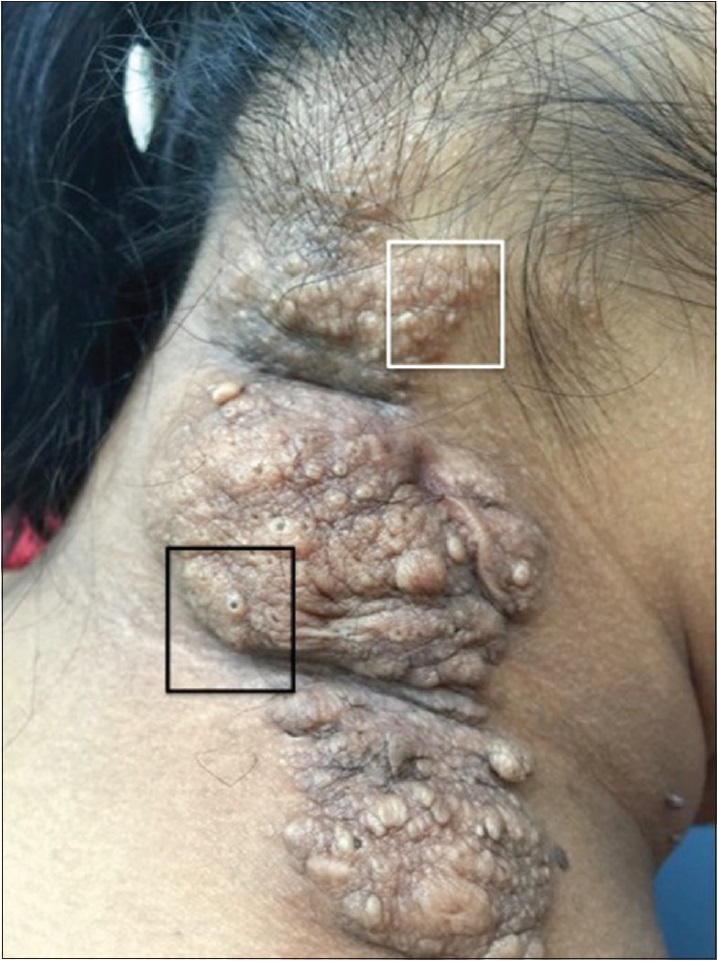 |
| Figure 1a: Nevus lipomatosus cutaneous superficialis on the nape of the neck. Dermatoscopic evaluation was conducted from sites highlighted by white and black boxes |
 |
| Figure 1b: Lobules and single scattered mature adipocytes in the upper dermis with overlying acanthosis of the epidermis (H and E, ×100) |
Dermatoscopic evaluation was performed in the index case from the sites marked by white [Figure 2a] and black [Figure 2b] boxes and two other patients [Figure 2c] and [Figure 2d] of biopsy proven nevus lipomatosus cutaneous superficialis using a DermLite II hybrid m dermatoscope at 10× magnification in polarized contact mode. Photographs were captured by Apple iPhone 6 Plus with the aid of DermLite app. Five dermatoscopic features were noted (1) cerebriform surface formed by gyri (ridges) and sulci (fissures), the latter filled with keratin products [Figure 2b], [Figure 2c] and [Figure 2d] (2) web-like regular pigment network consisting of brown lines and yellowish holes creating a honeycomb-like pattern [Figure 2c] and [Figure 2d] (3) rim of the cerebriform surface showed a “ground glass” white film or “veil” [Figure 2c], green arrow and arc] (4) yellowish structureless areas, some of them showing a perifollicular distribution [[Figure 2a] and [Figure 2d], red circles] and (5) comedo-like openings [[Figure 2b], black arrow].
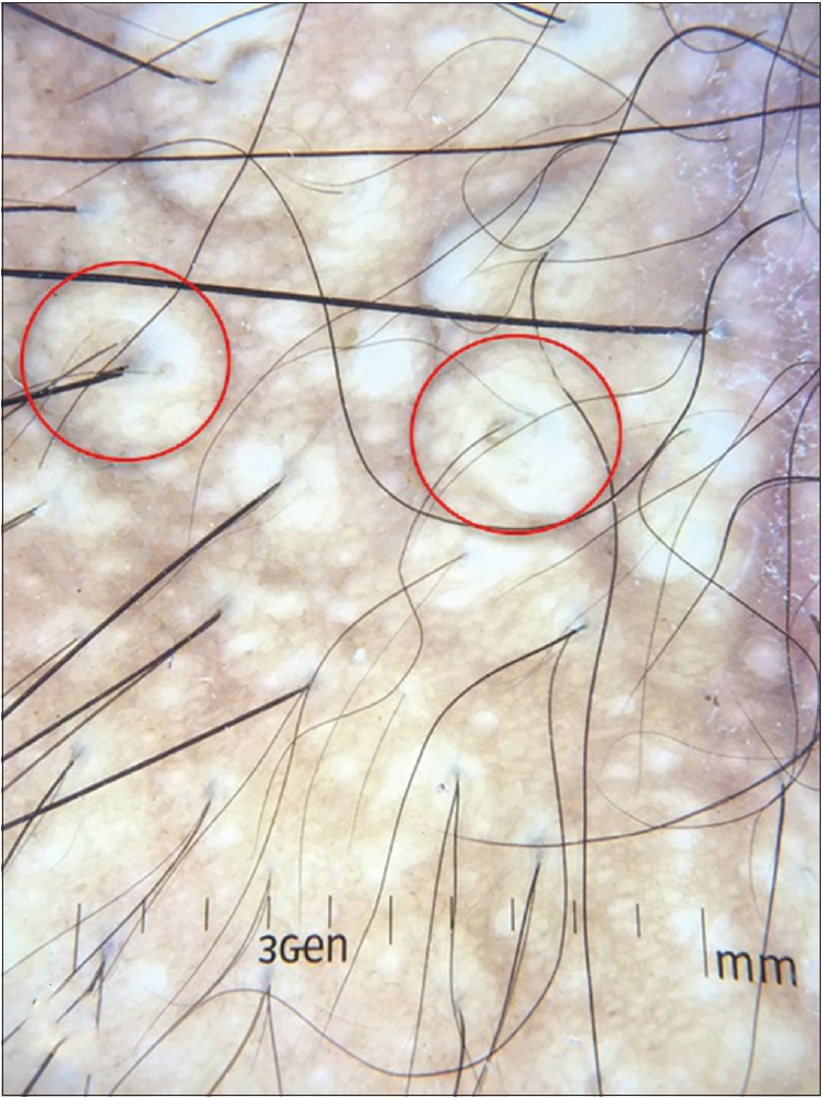 |
| Figure 2a: Dermatoscopic evaluation showing yellowish structureless areas (red circles), some of them in perifollicular distribution |
 |
| Figure 2b: Comedo-like opening (arrow) |
 |
| Figure 2c: Cerebriform surface with honeycomb-like pigmentary network admixed with yellowish holes and a rim of white veil (green arc and arrowhead) |
 |
| Figure 2d: Gyri and sulci with sulci containing keratinous debris and yellow structureless area (red circle) |
Two variants of nevus lipomatosus cutaneous superficialis have been described, the classical and the solitary.[2] The classic variant manifests as multiple, soft, pedunculated, cerebriform papules or nodules that often coalesce to form plaques in a zonal pattern. The common sites of occurrence of the classical variant are the flank, buttocks and upper thigh, though it is known to occur on unusual sites such as upper trunk, abdomen, axillae, genitalia and face where it can pose a diagnostic challenge.[3],[4] The solitary variant presents as a solitary, dome-shaped, sessile papule or nodule and can be misdiagnosed as giant skin tag or solitary neurofibroma. In such a situation, dermatoscopic evaluation aids in establishing the diagnosis and avoids skin biopsy, especially in infants and young children. However, we were unable to find any previous report describing the dermatoscopic features of nevus lipomatosus cutaneous superficialis.
The index case had her lesion located on the nape of the neck, an uncommon site for nevus lipomatosus cutaneous superficialis. The site and distribution of the lesion were suggestive of segmental neurofibromatosis and nevus sebaceous. Based on our experience with dermatoscopic evaluation of previous two cases, nevus lipomatosus cutaneous superficialis was strongly considered and was further confirmed by histopathological examination.
Dermatoscopic features of neurofibromatosis have been recently described and include peripheral pigmented network, a halo of brown pigmentation, pink-red structureless areas, fingerprint-like structures, scar-like areas, fissures and blood vessels.[5] Dermatoscopy of nevus sebaceous shows bright yellow dots not associated with hair follicles.[6] None of these features were seen in our patients.
The cerebriform surface seen on dermatoscopy represents the uneven surface seen clinically. The cerebriform surface formed by gyri and sulci may also be seen in dermal nevus and seborrheic keratosis while comedo-like openings are usually characteristic of seborrheic keratosis.[7] However, the presence of yellow structureless areas representing dermal adipocytes is more suggestive of nevus lipomatosus cutaneous superficialis. The presence of a regular pigment network in our cases indicates the relative histological preservation of the normal rete ridge in most nevus lipomatosus cutaneous superficialis as opposed to disrupted rete ridge anatomy in neurofibromas (epidermal flattening) and nevus sebaceous (acanthosis). The pigment network was distinct in our cases since all three patients belonged to skin phototype IV and V. This particular feature may require enhancement with nonpolarized immersion dermatoscopy in lower skin types. Yellow dots seen in nevus sebaceous represent sebaceous glands and are generally not associated with hair follicles in infancy.[6] However, the developed sebaceous glands during late childhood in nevus sebaceous may surround small hair follicles. Therefore, the likelihood of finding perifollicular yellow dots in nevus sebaceous is low (especially in infancy). In contrast, dermatoscopy of nevus lipomatosus cutaneous superficialis shows yellow structureless areas in perifollicular distribution around terminal hair follicles [Figure 2a].
In conclusion, no single dermatoscopic feature is distinctive of nevus lipomatosus cutaneous superficialis. However, the combination of the above-mentioned dermatoscopic features aids in differentiating it from neurofibroma and nevus sebaceous and is a helpful adjunct in its clinical diagnosis.
Financial support and sponsorship
Nil.
Conflicts of interest
There are no conflicts of interest.
| 1. | Russo T, Piccolo V, Lallas A, Argenziano G. Recent advances in dermoscopy. F1000Res 2016;5. pii: F1000Rev-184. [Google Scholar] |
| 2. | Moss C, Shahidullah H. Naevi and other Developmental Defects. In: Burns T, Breathnach S, Cox NH, Griffiths C, editors. Rook's Textbook of Dermatology. 8th ed. Oxford: Blackwell Scientific Publications; 2010. p. 658-9. [Google Scholar] |
| 3. | Lane JE, Clark E, Marzec T. Nevus lipomatosus cutaneus superficialis. Pediatr Dermatol 2003;20:313-4. [Google Scholar] |
| 4. | Sendhil Kumaran M, Narang T, Dogra S, Saikia UN, Kanwar AJ. Nevus lipomatosus superficialis unseen or unrecognized: A report of eight cases. J Cutan Med Surg 2013;17:335-9. [Google Scholar] |
| 5. | Duman N, Elmas M. Dermoscopy of cutaneous neurofibromas associated with neurofibromatosis type 1. J Am Acad Dermatol 2015;73:529-31. [Google Scholar] |
| 6. | Neri I, Savoia F, Giacomini F, Raone B, Aprile S, Patrizi A. Usefulness of dermatoscopy for the early diagnosis of sebaceous naevus and differentiation from aplasia cutis congenita. Clin Exp Dermatol 2009;34:e50-2. [Google Scholar] |
| 7. | Hirokawa D, Lee JB. Dermatoscopy: An overview of subsurface morphology. Clin Dermatol 2011;29:557-65. [Google Scholar] |
Fulltext Views
2,577
PDF downloads
907


