Generic selectors
Exact matches only
Search in title
Search in content
Post Type Selectors
Search in posts
Search in pages
Filter by Categories
15th National Conference of the IAOMFP, Chennai, 2006
Abstract
Abstracts from current literature
Acne in India: Guidelines for management - IAA Consensus Document
Addendum
Announcement
Art & Psychiatry
Article
Articles
Association Activities
Association Notes
Award Article
Book Review
Brief Report
Case Analysis
Case Letter
Case Letters
Case Notes
Case Report
Case Reports
Clinical and Laboratory Investigations
Clinical Article
Clinical Studies
Clinical Study
Commentary
Conference Oration
Conference Summary
Continuing Medical Education
Correspondence
Corrigendum
Cosmetic Dermatology
Cosmetology
Current Best Evidence
Current Issue
Current View
Derma Quest
Dermato Surgery
Dermatopathology
Dermatosurgery Specials
Dispensing Pearl
Do you know?
Drug Dialogues
e-IJDVL
Editor Speaks
Editorial
Editorial Remarks
Editorial Report
Editorial Report - 2007
Editorial report for 2004-2005
Errata
Erratum
Focus
Fourth All India Conference Programme
From Our Book Shelf
From the Desk of Chief Editor
General
Get Set for Net
Get set for the net
Guest Article
Guest Editorial
History
How I Manage?
IADVL Announcement
IADVL Announcements
IJDVL Awards
IJDVL AWARDS 2015
IJDVL Awards 2018
IJDVL Awards 2019
IJDVL Awards 2020
IJDVL International Awards 2018
Images in Clinical Practice
In Memorium
Inaugural Address
Index
Knowledge From World Contemporaries
Leprosy Section
Letter in Response to Previous Publication
Letter to Editor
Letter to the Editor
Letter to the Editor - Case Letter
Letter to the Editor - Letter in Response to Published Article
LETTER TO THE EDITOR - LETTERS IN RESPONSE TO PUBLISHED ARTICLES
Letter to the Editor - Observation Letter
Letter to the Editor - Study Letter
Letter to the Editor - Therapy Letter
Letter to the Editor: Articles in Response to Previously Published Articles
Letters in Response to Previous Publication
Letters to the Editor
Letters to the Editor - Letter in Response to Previously Published Articles
Letters to the Editor: Case Letters
Letters to the Editor: Letters in Response to Previously Published Articles
Medicolegal Window
Messages
Miscellaneous Letter
Musings
Net Case
Net case report
Net Image
Net Images
Net Letter
Net Quiz
Net Study
New Preparations
News
News & Views
Obituary
Observation Letter
Observation Letters
Oration
Original Article
ORIGINAL CONTRIBUTION
Original Contributions
Pattern of Skin Diseases
Pearls
Pediatric Dermatology
Pediatric Rounds
Perspective
Presedential Address
Presidential Address
Presidents Remarks
Quiz
Recommendations
Regret
Report
Report of chief editor
Report of Hon : Treasurer IADVL
Report of Hon. General Secretary IADVL
Research Methdology
Research Methodology
Resident page
Resident's Page
Resident’s Page
Residents' Corner
Residents' Corner
Residents' Page
Retraction
Review
Review Article
Review Articles
Reviewers 2022
Revision Corner
Self Assessment Programme
SEMINAR
Seminar: Chronic Arsenicosis in India
Seminar: HIV Infection
Short Communication
Short Communications
Short Report
Snippets
Special Article
Specialty Interface
Studies
Study Letter
Study Letters
Supplement-Photoprotection
Supplement-Psoriasis
Symposium - Contact Dermatitis
Symposium - Lasers
Symposium - Pediatric Dermatoses
Symposium - Psoriasis
Symposium - Vesicobullous Disorders
SYMPOSIUM - VITILIGO
Symposium Aesthetic Surgery
Symposium Dermatopathology
Symposium-Hair Disorders
Symposium-Nails Part I
Symposium-Nails-Part II
Systematic Review and Meta-Analysis
Systematic Reviews and Meta-analyses
Systematic Reviews and Meta-analysis
Tables
Technology
Therapeutic Guideline-IADVL
Therapeutic Guidelines
Therapeutic Guidelines - IADVL
Therapeutics
Therapy
Therapy Letter
Therapy Letters
View Point
Viewpoint
What’s new in Dermatology
View/Download PDF

Translate this page into:

Letter to the Editor - Case Letter
2016:82:3;330-332
doi: 10.4103/0378-6323.174384
PMID: 27088946

Verrucous oral focal mucinosis

Kinjal Deepak Rambhia, Uday Sharadchandra Khopkar
 Department of Dermatology, Seth GS Medical College and KEM Hospital, Mumbai, Maharashtra, India

Correspondence Address:
Kinjal Deepak Rambhia
B-105, Kalpataru Classic, Chincholi Bunder Road, Malad West, Mumbai - 400 064, Maharashtra
India
How to cite this article:
Rambhia KD, Khopkar US. Verrucous oral focal mucinosis. Indian J Dermatol Venereol Leprol 2016;82:330-332
Copyright: (C)2016 Indian Journal of Dermatology, Venereology, and Leprology

Sir,

A 60-year-old woman presented with an asymptomatic verrucous lesion on the right buccal mucosa that had grown slowly over the past 6 months. There was no history of bleeding from the lesion. The patient was a tobacco-chewer for many years. On examination, there was a cluster of verrucous papules coalesced to form a finger-like projection [Figure - 1]. The lesion was grayish-white, firm, non-tender and there was no bleeding to touch. The underlying and surrounding mucosa appeared normal. There was no cervical lymphadenopathy. General and systemic examinations were within normal limits.

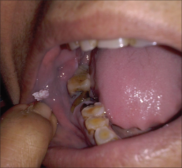
Figure 1: Clustered verrucous papules coalescing to form a plaque with finger-like projections

Biopsy from the lesion revealed a sub-epithelial, localized non-encapsulated area of loose myxomatous connective tissue stroma with stellate-shaped fibroblasts [Figure - 2] and [Figure - 3]. The overlying epidermis showed hyperkeratosis, acanthosis and papillomatosis. Alcian blue stain revealed blue staining in the areas of prominent mucin deposition [Figure - 4]. After clinico-pathological correlation, a diagnosis of verrucous focal mucinosis was made. The lesion was excised surgically.

Figure 2: Papillomatous projection with sub-epithelial bluish material (H and E, ×100)
Figure 3: Subepithelial mucin deposition (H and E, ×400)
Figure 4: Mucin deposition (Alcian blue, ×100)

Oral focal mucinosis is a rare, benign condition of unknown etiology. The first description was given by Tomich (1974) who reported eight such cases as the oral counterpart of cutaneous focal mucinosis.[1] It has been postulated that idiopathic focal hyperproduction of hyaluronic acid from the fibroblasts contributes to the pathogenesis. Oral focal mucinosis occurs primarily in adult patients and has a female preponderance. Oral mucosa overlying the bone such as gingiva and hard palate are most frequently involved, the next common areas of occurrence are buccal mucosa and tongue.[2] The classical clinical presentation of the lesion is as an asymptomatic, soft to firm, sessile or pedunculated mass with a smooth surface. The color of the lesion is the same as that of surrounding normal mucosa. It is most often mistaken for fibroma, pyogenic granuloma or mucocele. A smooth and non-ulcerated surface is the differentiating feature; however, a pebbled and lobulated surface has also been described.[2] In this case, the lesion was verrucous with grayish-white clusters of firm papules and normal surrounding mucosa. Clinical differential diagnoses of verrucous oral lesions include oral papilloma, verruca vulgaris, proliferating verrucous leukoplakia, verruciform xanthoma, verrucous carcinoma and papillary squamous cell carcinoma.[3] In the absence of distinct and specific clinical features, diagnosis is based on histopathology.

Oral papilloma and verruca vulgaris are characterized by hypergranulosis and koilocytes on biopsy. Proliferating verrucous leukoplakia presents with a hyperplastic epidermis and dysplasia. Verrucous carcinomas are locally aggressive low-grade malignancies with, koilocytes, individual cell keratinization and squamous pearl formation.[4] Papillary squamous cell carcinomas show large atypical cells with mitotic figures involving the entire epidermis. Histopathologically, verruciform xanthoma is characterized by foamy histiocytes in the papillary dermis. Oral focal mucinosis is characterized by a localized, sub-epithelial, non-encapsulated area of loose, myxomatous connective tissue encircled by normal collagen bundles. The myxomatous areas show minimal to absent reticulin fibers and fragmented collagen fibers replaced by mucin.[5] There are delicate fibrillary processes extending from the fibroblast cytoplasm. In superficial lesions, there is secondary atrophy of the epidermis with loss of rete ridges. In our patient, there was hyperkeratosis and papillomatosis overlying the sub-epithelial areas of localized mucin deposition which differs from previous reports. The histopathological differential diagnoses include soft-tissue myxoma, myxomatous change in fibrous lesions, nerve sheath myxoma and mucous retention phenomenon.[5] A sharp delineation and paucity of reticulin fibers differentiate oral focal mucinosis from soft tissue myxoma and myxomatous changes in fibrous lesions. Nerve sheath myxoma can be recognized by the presence of mast cells and a conspicuously lobular architecture. The mucous retention phenomenon or mucous retention cyst shows a circumscribed cavity containing mucoid material. The composition of the cavity is variable; it is formed by a lining of flattened cuboidal or columnar epithelial cells of the salivary gland duct in mucous retention cyst or compressed fibrous connective tissue or granulation tissue in mucous extravasation cyst. Infiltration by leukocytes and mononuclear phagocytes may be seen. In cases with atypical clinical features, histopathology helps to arrive at the correct diagnosis. The lesion is treated by surgical excision and has no tendency to recur.[6]

Financial support and sponsorship

Nil.

Conflicts of interest

There are no conflicts of interest.

References
1.
Tomich CE. Oral focal mucinosis: A clinico pathologic and histochemical study of eight cases. Oral Surg 1974;38:714-24.
[Google Scholar]
2.
Madhusudhan AS, Nagarajappa D, Manjunatha BS, Swati S, Charan Babu HS. Oral focal mucinosis: Report of two cases. Rev Odonto Cienc 2010;25:310-3.
[Google Scholar]
3.
Swetha P, Supriya NA, Kumar GR. Characterization of different verrucous mucosal lesions. Indian J Dent Res 2013;24:642-4.
[Google Scholar]
4.
Terada T. Verrucous carcinoma of the oral cavity: A histopathologic study of 10 Japanese cases. J Maxillofac Oral Surg 2011;10:148-51.
[Google Scholar]
5.
Ena S, Nadella M, Chatterjee A, Ramesh A. Oral focal mucinosis: A rare case report of two cases. Ethiop J Health Sci 2013;23:178-82.
[Google Scholar]
6.
Bharti V, Singh J. Oral focal mucinosis of palatal mucosa: A rare case report. Contemp Clin Dent 2012;3:214-8.
[Google Scholar]

Fulltext Views
2,447

PDF downloads
1,752
Show Sections